. 2021 May 17;56(10):1484-1497.e7.
doi: 10.1016/j.devcel.2021.03.031. Epub 2021 Apr 19.

A G protein-coupled receptor-like module regulates cellulose synthase secretion from the endomembrane system in Arabidopsis

Affiliations

A G protein-coupled receptor-like module regulates cellulose synthase secretion from the endomembrane system in Arabidopsis

Heather E McFarlane et al. Dev Cell. .

Abstract

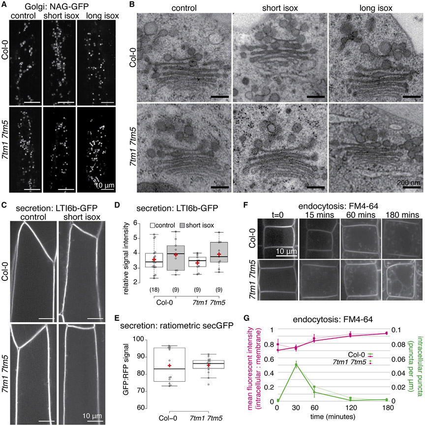
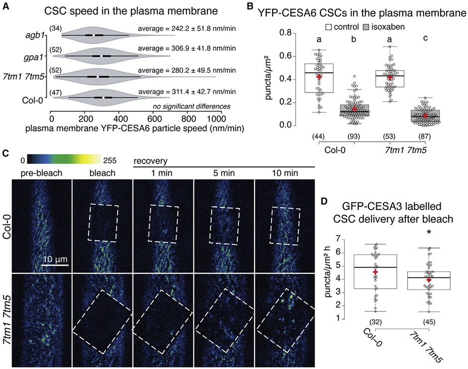
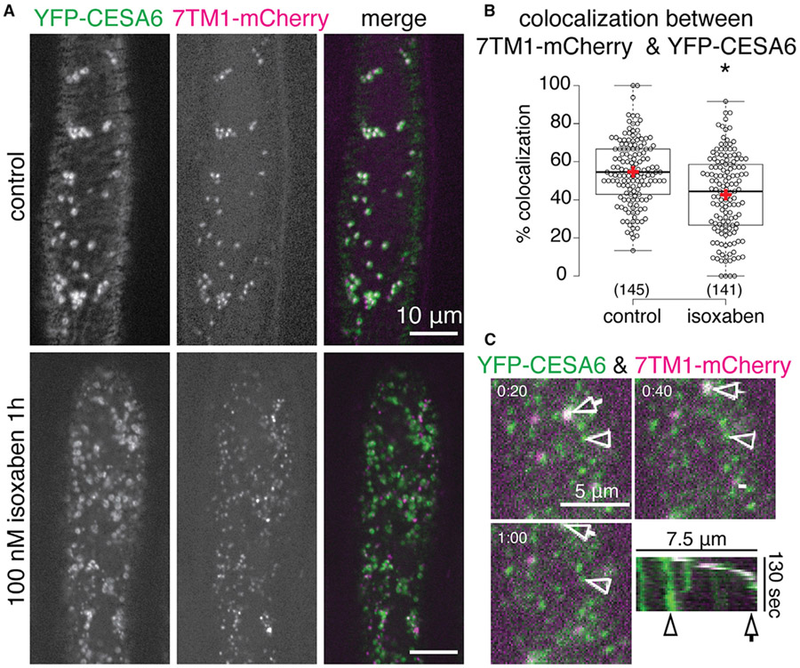
Cellulose is produced at the plasma membrane of plant cells by cellulose synthase (CESA) complexes (CSCs). CSCs are assembled in the endomembrane system and then trafficked to the plasma membrane. Because CESAs are only active in the plasma membrane, control of CSC secretion regulates cellulose synthesis. We identified members of a family of seven transmembrane domain-containing proteins (7TMs) that are important for cellulose production during cell wall integrity stress. 7TMs are often associated with guanine nucleotide-binding (G) protein signaling and we found that mutants affecting the Gβγ dimer phenocopied the 7tm mutants. Unexpectedly, the 7TMs localized to the Golgi/trans-Golgi network where they interacted with G protein components. Here, the 7TMs and Gβγ regulated CESA trafficking but did not affect general protein secretion. Our results outline how a G protein-coupled module regulates CESA trafficking and reveal that defects in this process lead to exacerbated responses to cell wall integrity stress.

Keywords: G protein-coupled receptor; GPCR; cell wall signaling; cellulose; cellulose synthesis; plant cell biology; plant cell walls; secretion.


Conflict of interest statement

Declaration of interests The authors declare no competing interests.

Figures

Figure 1.
Figure 1.. Mutations in 7TM family members cause increased sensitivity to cell wall stress
(A) Representative images of 7-day-old light-grown seedlings grown on control media and media supplemented with 2 nM isoxaben (isox). (B) Quantification of root lengths of seedlings represented in (A). (C) Representative images of etiolated 7-day-old seedlings grown on control media and media supplemented with 2 nM isoxaben. (D) Quantification of hypocotyl lengths of seedlings represented in (C). (E) Quantification of 7-day-old etiolated hypocotyl lengths of seedlings grown on control media and media supplemented with 150 nM DCB. (F) Cryo-scanning electron microscopy (SEM) of 3-day-old etiolated hypocotyls of seedlings grown on media supplemented with 2 nM isoxaben. (G) Quantification of cell length: width ratio from SEM images as a measure of cell swelling. (H) Quantification of cellulose content in different genotypes. All graphs summarize three independent experiments (two for SEM), n is indicated in parentheses; samples not sharing a common letter are significantly different (one-way ANOVA and Tukey HSD test, p < 0.05); in bar charts bars represent mean ± SE and in box plots box limits indicate 25th and 75th percentiles, whiskers extend to 1.5 times the interquartile range, median is indicated by a line, mean by a red “+” and individual data points are shown. Scale bars represent 5 mm in (A and C) and 500 or 50 mm as indicated in (F).
Figure 2.
Figure 2.. Mutations in components of the G protein signaling complex cause increased sensitivity to cell wall stress
(A) Representative images of 7-day-old light-grown seedlings grown on control media and media supplemented with 2 nM isoxaben. (B) Quantification of root lengths of seedlings represented in A. (C) Representative images of etiolated 7-day-old seedlings grown on control media and media supplemented with 2 nM isoxaben. (D) Quantification of hypocotyl lengths of seedlings represented in (B). (E) Cryo-scanning electron microscopy (SEM) of 3-day-old etiolated hypocotyls of seedlings grown on media supplemented with 2 nM isoxaben. (F) Quantification of cell length: width ratio from SEM images as a measure of cell swelling. (G) Quantification of cellulose content in different genotypes. All graphs summarize three independent experiments (two for SEM), Col-0 data in (B, D, and F) are the same as in Figure 1 since all samples were grown together, n is indicated in parentheses; samples not sharing a common letter are significantly different (one-way ANOVA and Tukey HSD test, p < 0.05); in bar charts bars represent mean ± SE and in box plots box limits indicate 25th and 75th percentiles, whiskers extend to 1.5 times the interquartile range, median is indicated by a line, mean by a red “+” and individual data points are shown. Scale bars represent 5 mm in (A and C) and 500 or 50 μm as indicated in (E).
Figure 3.
Figure 3.. 7TMs interact with components of the G protein complex
(A) Quantification of etiolated hypocotyl lengths of combinatorial mutants between the 7tm mutants and Gβ mutant agb1-2. (B) Bimolecular fluorescence complementation (BiFC) assay in transiently infiltrated Nicotiana benthamiana leaf epidermal cells with the BiFC signal shown in green and Golgi marker (XYLT) in magenta in the Merge & Zoom column; white boxes indicate the zoom region. Experiment was repeated three times with similar results. In box plot, box limits indicate 25th and 75th percentiles, whiskers extend to 1.5 times the interquartile range, median is indicated by a line, mean by a red “+”, individual data points are shown, n is indicated in parentheses and samples not sharing a common letter are significantly different (one-way ANOVA and Tukey HSD test, p < 0.05). Scale bars represent 10 or 2 μm as indicated in (B).
Figure 4.
Figure 4.. The 7TMs are localized to the Golgi apparatus and trans-Golgi network
(A) Functional 7TM1-YFP (native promoter-driven genomic 7TM1-3xYFP) and 7TM5-CFP (35S-driven) fusion proteins localize to intracellular compartments in 3-day-old etiolated hypocotyl cells. (B) Representative images from colocalization experiments between 7TM1-3xYFP (green) and 7TM5-CFP (magenta) or between 7TM1-3xYFP or 7TM5-CFP (green) and different endomembrane markers (magenta) as indicated in 3-day-old seedling root cells. C. Quantification of percent colocalization between 7TM1-3xYFP, 7TM1-mCherry, or 7TM5-CFP and endomembrane system markers. Graph summarizes three independent experiments, n (cells, no more than three cells imaged per seedling) is indicated in parentheses, bars represent mean ± SE Scale bars represent 10 μm in (A) and 2 μm in (B).
Figure 5.
Figure 5.. Overall Golgi structure and function are unaffected in 7tm mutants
(A) Representative images of the Golgi marker, NAG-GFP in 3-day-old wild type and 7tm1 7tm5 mutant etiolated hypocotyl cells after short-term (100 nM for 1 h) or long-term (2 nM for 3 days) isoxaben treatment, compared with control. (B) Representative images of the Golgi apparatus in high-pressure frozen, freeze-substituted 3-day-old wild type and 7tm1 7tm5 mutant etiolated hypocotyl cells after short-term or long-term isoxaben treatment, relative to control. (C) Representative images of the plasma membrane marker, GFP-LTI6b in 3-day-old wild type and 7tm1 7tm5 mutant etiolated hypocotyl cells after short-term isoxaben treatment. (D) Quantification of signal intensity from GFP-LTI6b signal represented in (C). (E) Quantification of ratiometric sec-GFP (35S:stN-RM-2A-sec-GFP) in 7tm1 7tm5 mutants, compared with wild type, in 3-day-old etiolated hypocotyls. (F) Representative time course images of membrane dye (FM4-64) uptake into in root cells of 3-day-old seedlings of 7tm1 7tm5 mutants, compared with wild type. FM4-64 initially labels the plasma membrane, then travels to the early endosome/TGN, late endosome and the tonoplast (vacuole membrane) over 3 h. (G) Quantification of FM4-64 uptake represented in (F). All graphs summarize three independent experiments, n (cells, no more than three cells imaged per seedling) is indicated in parentheses and there were no statistically significant differences between samples (one-way ANOVA and Tukey HSD test, p < 0.05). In box plots, box limits indicate 25th and 75th percentiles, whiskers extend to 1.5 times the interquartile range, median is indicated by a line, mean by a red “+” and individual data points are shown; in line plot, symbols represent mean ± SE. Scale bars represent 10 μm in (A, C, F, H) and 200 nm in (B).
Figure 6.
Figure 6.. 7tm mutants are defective in secretion of CSCS to the plasma membrane
(A) Quantification of CSC speeds in the plasma membrane of 3-day-old wild type and mutant etiolated hypocotyl cells. (B) Quantification of YFP-CESA6 labeled CSC particle density in the plasma membrane of 3-day-old etiolated hypocotyl cells in wild type versus 7tm1 7tm5 mutants after control or long-term isoxaben treatment (2 nM for 3 days). (C) Representative time course images of photobleaching of GFP-CESA3 in wild type and 7tm1 7tm5 mutant 3-day-old etiolated hypocotyl cells. Bleached area is indicated by dashed box, images are false-colored according to the scale indicated. (D) Quantification of the rate of CSC recovery in the plasma membrane after photobleaching in wild type versus 7tm1 7tm5 mutant 3-day-old etiolated hypocotyl cells. All graphs summarize three independent experiments, n (cells, no more than three cells imaged per seedling) is indicated in parentheses, samples not sharing a common letter in B are significantly different (one-way ANOVA and Tukey HSD test, p < 0.05) and in (D) samples marked by an × are significantly different from control (one-way t test, p < 0.05). In violin plot, white circles show the medians, wide bar limits indicate the 25th and 75th percentiles, whiskers extend 1.5 times the interquartile range, polygons represent density estimates of data and extend to extreme values. In box plot, box limits indicate 25th and 75th percentiles, whiskers extend to 1.5 times the interquartile range, median is indicated by a line, mean by a red “+” and individual data points are shown. Scale bars represent 10 μm in (C).
Figure 7.
Figure 7.. 7TM1 localization is biased from Golgi/TGN to SmaCCs upon cell wall stress treatment
(A) Representative images of YFP-CESA6 and 7TM1-mCherry colocalization in 3-day-old etiolated hypocotyl cells after control or short-term (100 nM for 1 h) isoxaben treatment. (B) Quantification of percent colocalization between YFP-CESA6 and 7TM1-mCherry in 3-day-old etiolated hypocotyl cells after control or short-term isoxaben treatment. C. Representative time-lapse images of YFP-CESA6 and 7TM1-mCherry colocalization in 3-day-old etiolated hypocotyl cells after control or short-term isoxaben treatment and kymograph indicating dynamics of YGP-CESA6 and 7TM1-mCherry labeled particles; timestamp is in minutes:seconds. Graph summarizes three independent experiments, n (cells, no more than three cells imaged per seedling) is indicated in parentheses, samples marked by an × are significantly different from control (one-way t test, p < 0.05), box limits indicate 25th and 75th percentiles, whiskers extend to 1.5 times the interquartile range, median is indicated by a line, mean by a red “+” and individual data points are shown. Scale bars represent 10 μm in (A) and 5 μm or 7.5 μm as indicated in (C).

Similar articles

Cited by

References

    1. Amsbury S, Hunt L, Elhaddad N, Baillie A, Lundgren M, Verhertbruggen Y, Scheller HV, Knox JP, Fleming AJ, and Gray JE (2016). Stomatal function requires pectin de-methyl-esterification of the guard cell wall. Curr. Biol 26, 2899–2906. - PMC - PubMed
    1. Anderson CT, and Kieber JJ (2020). Dynamic Construction, perception, and remodeling of plant cell walls. Annu. Rev. Plant Biol 71, 39–69. - PubMed
    1. Anderson DJ, and Botella JR (2007). Expression analysis and subcellular localization of the Arabidopsis thaliana G-protein beta-subunit AGB1. Plant Cell Rep. 26, 1469–1480. - PubMed
    1. Aranda-Sicilia MN, Trusov Y, Maruta N, Chakravorty D, Zhang Y, and Botella JR (2015). Heterotrimeric G proteins interact with defense-related receptor-like kinases in Arabidopsis. J. Plant Physiol 188, 44–48. - PubMed
    1. Arioli T, Peng L, Betzner AS, Burn J, Wittke W, Herth W, Camilleri C, Höfte H, Plazinski J, Birch R, et al. (1998). Molecular analysis of cellulose biosynthesis in Arabidopsis. Science 279, 717–720. - PubMed

Publication types

MeSH terms

LinkOut - more resources