. 2021 Apr 20;13(8):11786-11807.
doi: 10.18632/aging.202876. Epub 2021 Apr 20.

Identification of prognostic biomarkers associated with the occurrence of portal vein tumor thrombus in hepatocellular carcinoma

Affiliations

Identification of prognostic biomarkers associated with the occurrence of portal vein tumor thrombus in hepatocellular carcinoma

Tong Lin et al. Aging (Albany NY). .

Abstract

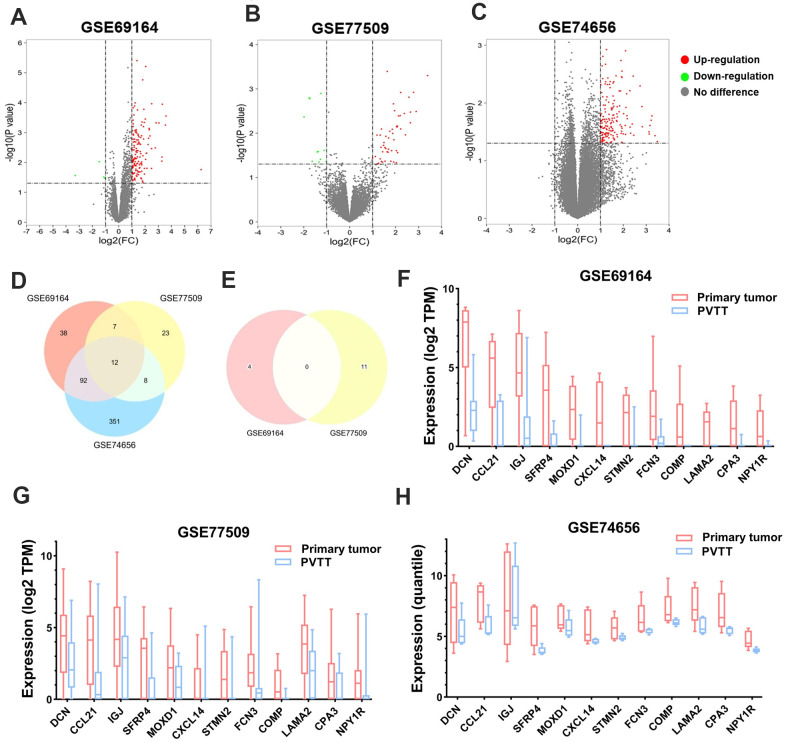
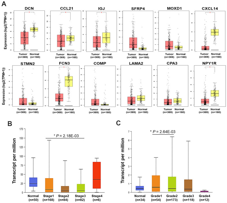
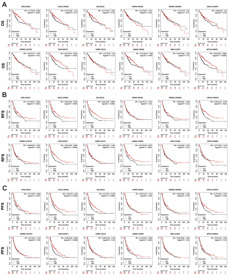
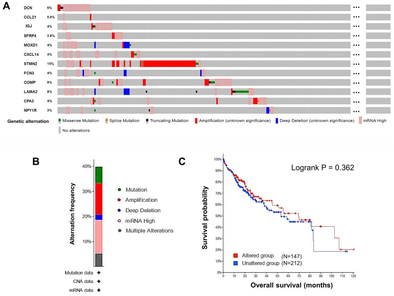
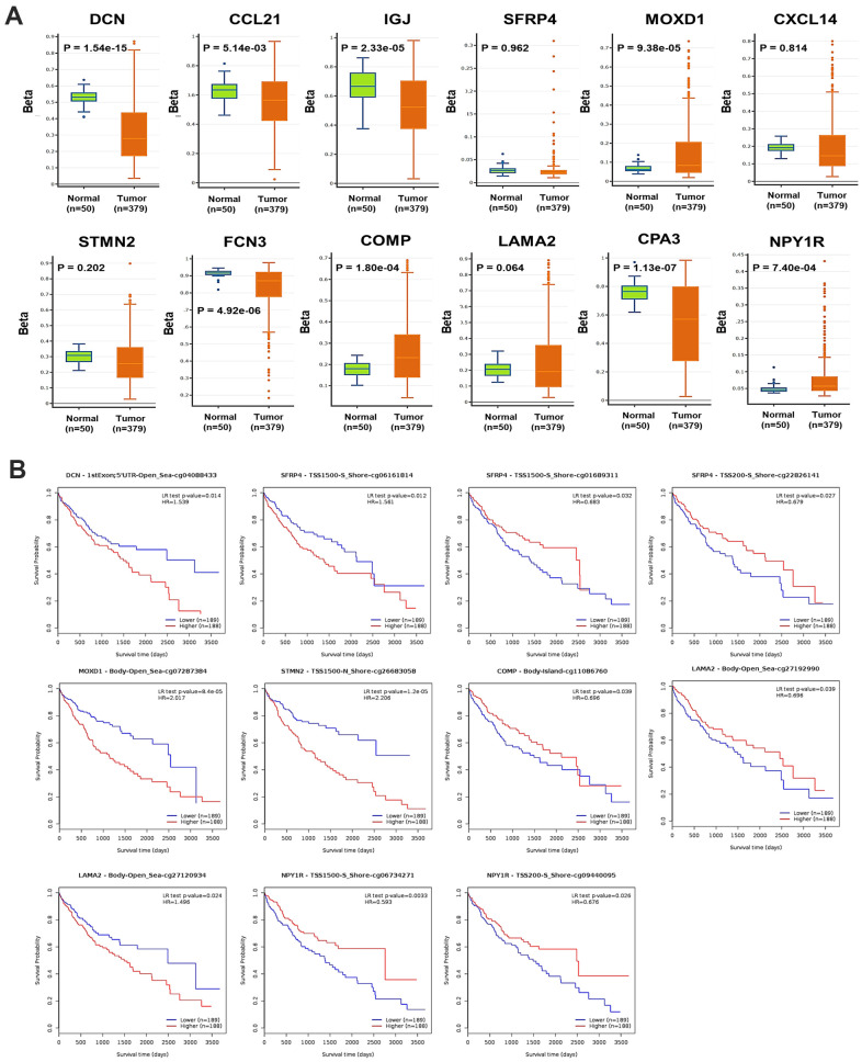
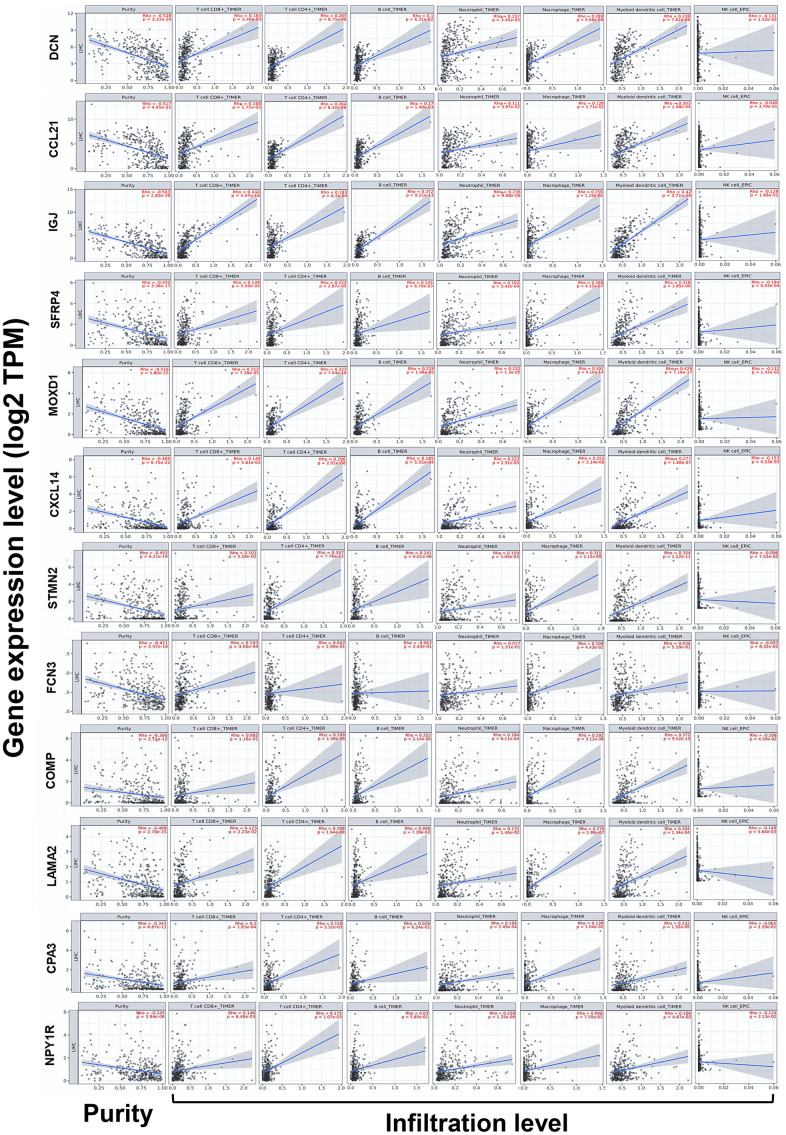
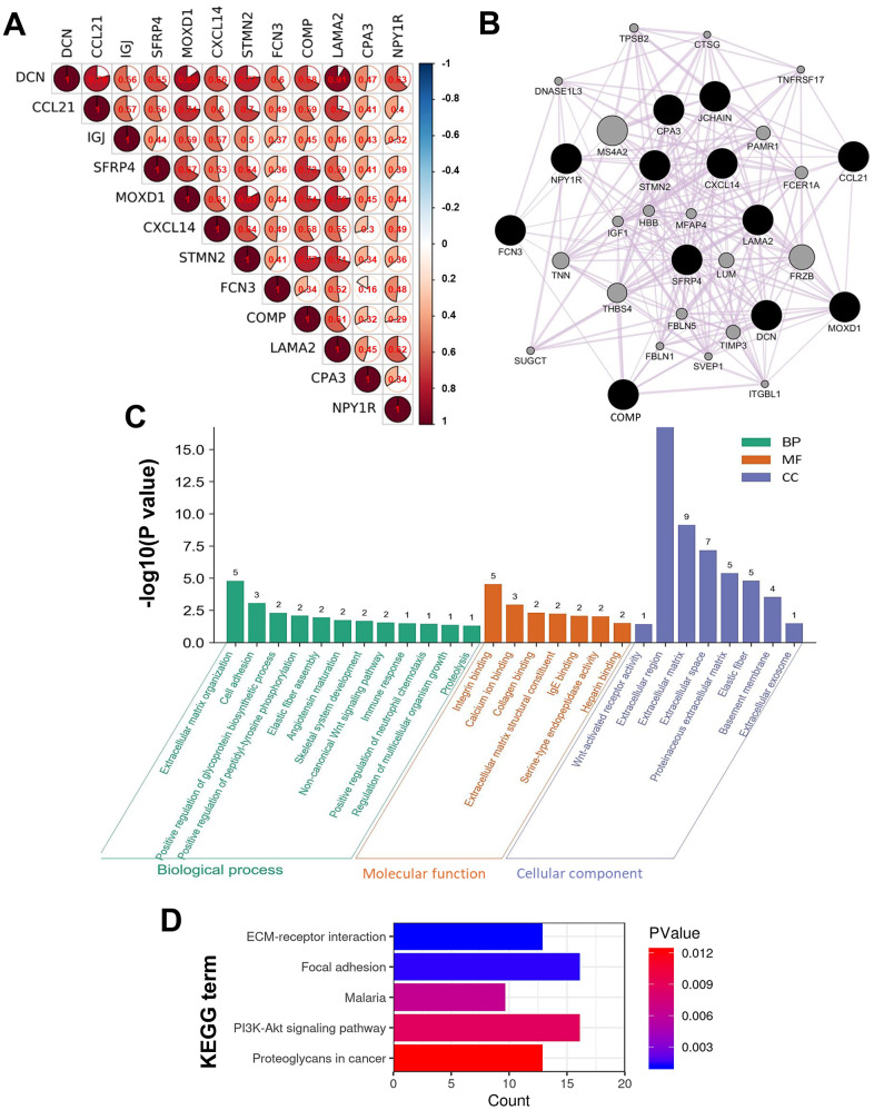
The occurrence of portal vein tumor thrombus (PVTT) is strongly correlated to the staging and poor prognosis of hepatocellular carcinoma (HCC) patients. However, the mechanisms of PVTT formation remain unclear. This study aimed to investigate differentially expressed genes (DEGs) between primary tumor (PT) and PVTT tissues and comprehensively explored the underlying mechanisms of PVTT formation. The DEGs between PT and paired PVTT tissues were analyzed using transcriptional data from the Gene Expression Omnibus (GEO) database. The expression, clinical relevance, prognostic significance, genetic alternations, DNA methylation, correlations with immune infiltration, co-expression correlations, and functional enrichment analysis of the DEGs were explored using multiple databases. As result, 12 DEGs were commonly down-expressed in PVTT compared with PT tissues among three datasets. The expression of DCN, CCL21, IGJ, CXCL14, FCN3, LAMA2, and NPY1R was progressively decreased from normal liver, PT, to PVTT tissues, whose up-expression associated with favorable survivals of HCC patients. The genetic alternations and DNA methylation of the DEGs frequently occurred, and several methylated CpG sites of the DEGs significantly correlated with outcomes of HCC patients. The immune infiltration in the tumor microenvironment of HCC was correlated with the expression level of the DEGs. Besides, the DEGs and their co-expressive genes participated in the biological processes of extracellular matrix (ECM) organization and focal adhesion. In summary, this study indicated the dysregulation of ECM and focal adhesion might contribute to the formation of PVTT. And the above seven genes might serve as potential biomarkers of PVTT occurrence and prognosis of HCC patients.

Keywords: bioinformatics; hepatocellular carcinoma; immune infiltration; portal vein tumor thrombus; prognosis.


Conflict of interest statement

CONFLICTS OF INTEREST: The authors declare that they have no conflicts of interest.

Figures

Figure 1
Figure 1
Identification of the DEGs between PT and PVTT tissues. Volcano plots showing the identification of the DEGs in (A) GSE69164, (B) GSE77509, and (C) GSE74656 datasets with the screening criteria of |log2(FC)| > 1 and P < 0.05. Dots in red or green represent upregulated or downregulated DEGs in PT compared with PVTT tissues, dots in grey represent genes without significant expressional differences. Venn diagrams showing the intersections of (D) upregulated and (E) downregulated DEGs among the three datasets. Since no downregulated DEGs was identified in dataset GSE74656, the Venn diagram for it was not drawn. The expression of the 12 overlapping upregulated DEGs in PT and PVTT tissues in datasets (F) GSE69164, (G) GSE77509, and (H) GSE74656. DEGs, differentially expressed genes; PT, primary tumor; PVTT, portal vein tumor thrombus; FC, fold change.
Figure 2
Figure 2
Expression and clinical relevance of the DEGs. (A) The expression of the 12 DEGs in HCC and normal liver samples (GEPIA) (*P < 0.05). (B) The expression of CCL21 in HCC and normal liver samples by different stages. (C) The expression of MOXD1 in HCC and normal liver samples by different grades. TPM, transcript per million.
Figure 3
Figure 3
Prognostic significance of the DEGs in HCC patients (KM Plotter). The survival curves showed the associations between the expression of the 12 DEGs with (A) OS, (B) RFS, and (C) PFS of HCC patients. OS, overall survival; RFS, relapse free survival; PFS, progression free survival; HR, hazard ratio.
Figure 4
Figure 4
Genetic alternations of the DEGs in HCC patients (cBioPortal). (A) The overview of the genetic alternations occurring in the 12 DEGs in HCC patients from “TCGA, Firehose Legacy” dataset. (B) The summary graph of alternation frequency of the 12 DEGs in HCC patients. (C) The effect of the overall genetic alternations of the DEGs on OS of HCC patients.
Figure 5
Figure 5
DNA methylation of the DEGs in HCC. (A) The global DNA methylation level of the 12 DEGs in HCC and normal liver samples (DNMIVD). (B) The associations between the methylation level of CpG sites of the DEGs with the OS of HCC patients (MethSurv).
Figure 6
Figure 6
Correlations between the expression of the DEGs with immune infiltration in HCC (TIMER). Correlations between the expression of the 12 DEGs with tumor purity, and infiltration level of CD8+ T cells, CD4+ T cells, B cells, neutrophils, macrophages, DCs, and NK cells in HCC. DCs, dendritic cells; NK cells, natural killer cells.
Figure 7
Figure 7
Intergenic correlations, co-expression network, and the biological functions of the DEGs. (A) Intergenic correlations of the 12 DEGs. (B) The co-expression network of the 12 DEGs constructed by GeneMANIA. Edges in the network represent the co-expression relations among genes. The results of (C) GO and (D) KEGG functional enrichment analyses for all the genes in the co-expression network.

References

    1. Bray F, Ferlay J, Soerjomataram I, Siegel RL, Torre LA, Jemal A. Global cancer statistics 2018: GLOBOCAN estimates of incidence and mortality worldwide for 36 cancers in 185 countries. CA Cancer J Clin. 2018; 68:394–424. 10.3322/caac.21492 - DOI - PubMed
    1. Duran SR, Jaquiss RD. Hepatocellular Carcinoma. N Engl J Med. 2019; 381:e2. 10.1056/NEJMc1906565 - DOI - PubMed
    1. Cheng S, Chen M, Cai J, Sun J, Guo R, Bi X, Lau WY, Wu M. Chinese expert consensus on multidisciplinary diagnosis and treatment of hepatocellular carcinoma with portal vein tumor thrombus (2018 edition). Liver Cancer. 2020; 9:28–40. 10.1159/000503685 - DOI - PMC - PubMed
    1. Lu J, Zhang XP, Zhong BY, Lau WY, Madoff DC, Davidson JC, Qi X, Cheng SQ, Teng GJ. Management of patients with hepatocellular carcinoma and portal vein tumour thrombosis: comparing east and west. Lancet Gastroenterol Hepatol. 2019; 4:721–30. 10.1016/S2468-1253(19)30178-5 - DOI - PubMed
    1. Chan SL, Chong CC, Chan AW, Poon DM, Chok KS. Management of hepatocellular carcinoma with portal vein tumor thrombosis: review and update at 2016. World J Gastroenterol. 2016; 22:7289–300. 10.3748/wjg.v22.i32.7289 - DOI - PMC - PubMed

MeSH terms

Substances