. 2021 Apr;11(4):210026.
doi: 10.1098/rsob.210026. Epub 2021 Apr 21.

Substrate recruitment by zDHHC protein acyltransferases

Affiliations

Substrate recruitment by zDHHC protein acyltransferases

Martin Ian P Malgapo et al. Open Biol. 2021 Apr.

Abstract

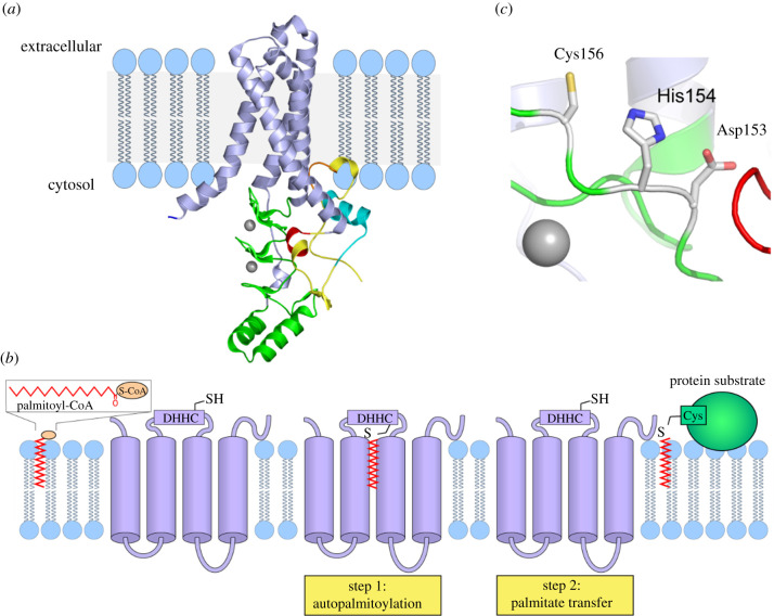
Protein palmitoylation is the post-translational attachment of fatty acids, most commonly palmitate (C16 : 0), onto a cysteine residue of a protein. This reaction is catalysed by a family of integral membrane proteins, the zDHHC protein acyltransferases (PATs), so-called due to the presence of an invariant Asp-His-His-Cys (DHHC) cysteine-rich domain harbouring the catalytic centre of the enzyme. Conserved throughout eukaryotes, the zDHHC PATs are encoded by multigene families and mediate palmitoylation of thousands of protein substrates. In humans, a number of zDHHC proteins are associated with human diseases, including intellectual disability, Huntington's disease, schizophrenia and cancer. Key to understanding the physiological and pathophysiological importance of individual zDHHC proteins is the identification of their protein substrates. Here, we will describe the approaches and challenges in assigning substrates for individual zDHHCs, highlighting key mechanisms that underlie substrate recruitment.

Keywords: fatty acylation; palmitoylation; post-translational modification; zDHHC enzyme.


Figures

Figure 1.
Figure 1.
Diverse membrane topology and conserved motifs in the zDHHC enzyme family. (a) Most zDHHC proteins contain four TMDs. The DHHC-CRD (dark green) harbours the canonical DHHC motif (light green) situated on the cytoplasmic face of the lipid bilayer between TMD 2 and 3. Additional conserved features within the family include the DPG (yellow), TTxE (red) and PaCCT (blue) motifs. (b) zDHHC13 and zDHHC17 are predicted to contain six TMDs with ankyrin repeats at the N-terminus. (c) Sequence analysis predicts that two zDHHC proteins contain five TMDs. In zDHHC24, this implies that the C-terminus faces the lumenal side of the lipid bilayer.
Figure 2.
Figure 2.
Phylogenetic analysis of human zDHHC PATs. (a) Phylogenetic tree of the 23 human zDHHCs PATs. The proteins were categorized into several subfamilies based on the homology of the full amino acid sequences using Clustal Omega software. (b) Human zDHHC PATs were grouped loosely to highlight differences in protein size and the presence of conserved binding domains and motifs.
Figure 3.
Figure 3.
Structure and kinetic mechanism of zDHHC-mediated protein palmitoylation. (a) Structure of human zDHHC20 (PDB entry 6BML [14]) represented with cartoons using Pymol. TMDs 1–4 are represented as blue helices. The DHHC-CRD is represented in green, C-terminal domain (CTD) in yellow. An amphipathic helix in the CTD is represented in orange. The TTxE motif is represented in red and the PaCCT motif in cyan. The two zinc ions are represented as grey spheres. (b) Using palmitoyl CoA as a donor molecule, the zDHHC enzyme first modifies the cysteine of the DHHC motif with palmitate, and then transfers the palmitate from itself to the protein substrate. (c) The side chains involved in the catalytic triad are represented as sticks and coloured by element (C-grey, N-blue, S-yellow, O-red).

References

    1. Chen B, Sun Y, Niu J, Jarugumilli GK, Wu X. 2018. Protein lipidation in cell signaling and diseases: function, regulation, and therapeutic opportunities. Cell Chem. Biol. 25, 817-831. (10.1016/j.chembiol.2018.05.003) - DOI - PMC - PubMed
    1. Jiang H, Zhang X, Chen X, Aramsangtienchai P, Tong Z, Lin H. 2018. Protein lipidation: occurrence, mechanisms, biological functions, and enabling technologies. Chem. Rev. 118, 919-988. (10.1021/acs.chemrev.6b00750) - DOI - PMC - PubMed
    1. Blanc M, David F, Abrami L, Migliozzi D, Armand F, Bürgi J, van der Goot F.. 2015. SwissPalm: protein palmitoylation database. F1000Research 4, 261. (10.12688/f1000research.6464.1) - DOI - PMC - PubMed
    1. Broncel M, Serwa RA, Ciepla P, Krause E, Dallman MJ, Magee AI, Tate EW. 2015. Multifunctional reagents for quantitative proteome-wide analysis of protein modification in human cells and dynamic profiling of protein lipidation during vertebrate development. Angew. Chem. Int. Ed. 54, 5948. (10.1002/anie.201500342) - DOI - PMC - PubMed
    1. Won SJ, Martin BR. 2018. Temporal profiling establishes a dynamic S-palmitoylation cycle. ACS Chem. Biol. 13, 1560-1568. (10.1021/acschembio.8b00157) - DOI - PMC - PubMed

Publication types

LinkOut - more resources