

The many faces of solitary fibrous tumor; diversity of histological features, differential diagnosis and role of molecular studies and surrogate markers in avoiding misdiagnosis and predicting the behavior
- PMID: 33879215
- PMCID: PMC8059036
- DOI: 10.1186/s13000-021-01095-2
The many faces of solitary fibrous tumor; diversity of histological features, differential diagnosis and role of molecular studies and surrogate markers in avoiding misdiagnosis and predicting the behavior
Abstract
Background: Solitary Fibrous Tumor (SFT) is a distinct soft tissue neoplasm associated with NAB2-STAT6 gene fusion. It can involve a number of anatomic sites and exhibits a wide spectrum of histological features.
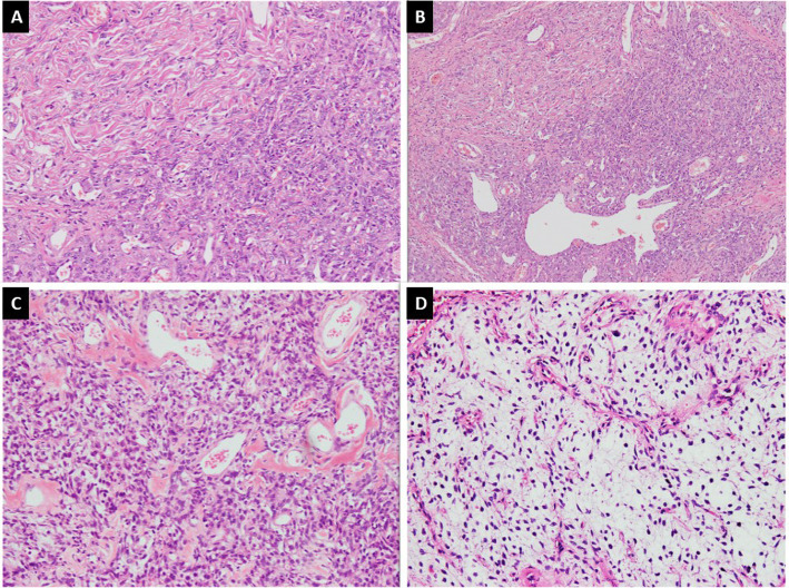
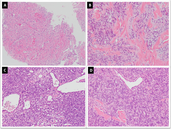
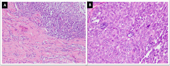
Main body: Apart from diversity in morphological features seen even in conventional SFT, two histologic variants (fat-forming and giant cell-rich) are also recognized. In addition, a malignant form and dedifferentiation are well recognized. Owing to diverse histological features and involvement of diverse anatomic locations, SFT can mimic other soft tissue neoplasms of different lineages including schwannoma, spindle cell lipoma, dermatofibrosarcoma protuberans, liposarcoma, gastrointestinal stromal tumor (GIST), malignant peripheral nerve sheath tumor (MPNST), and synovial sarcoma. SFT is classified as an intermediate (rarely metastasizing) tumor according to World Health Organization Classification of Tumors of Soft tissue and Bone, 5th edition. The management and prognosis of SFT differs from its malignant mimics and correct diagnosis is therefore important. Although SFT expresses a distinct immunohistochemical (IHC) profile, the classic histomorphological and IHC profile is not seen in all cases and diagnosis can be challenging. NAB2-STAT6 gene fusion has recently emerged as a sensitive and specific molecular marker and its IHC surrogate marker signal transducer and activator of transcription 6 (STAT6) has also shown significant sensitivity and specificity. However, few recent studies have reported STAT6 expression in other soft tissue neoplasms.
Conclusion: This review will focus on describing the diversity of histological features of SFT, differential diagnoses and discussing the features helpful in distinguishing SFT from its histological mimics.
Keywords: CD34; Hemangiopericytoma; Immunohistochemistry; NAB2-STAT6; STAT-6; Solitary fibrous tumor; Staghorn, fusion transcript.
Conflict of interest statement
The authors declare that there are no conflicts of interest.
Figures


Similar articles
-
Extrapleural solitary fibrous tumor: A distinct entity from pleural solitary fibrous tumor. An update on clinical, molecular and diagnostic features.Ann Diagn Pathol. 2018 Jun;34:142-150. doi: 10.1016/j.anndiagpath.2018.01.004. Epub 2018 Apr 9. Ann Diagn Pathol. 2018. PMID: 29660566 Review.
-
Molecular characterization of a series of solitary fibrous tumors, including immunohistochemical expression of STAT6 and NATB2-STAT6 fusion transcripts, using Reverse Transcriptase(RT)-Polymerase chain reaction(PCR) technique: An Indian experience.Pathol Res Pract. 2017 Nov;213(11):1404-1411. doi: 10.1016/j.prp.2017.08.011. Epub 2017 Aug 26. Pathol Res Pract. 2017. PMID: 28869107
-
Solitary fibrous tumors of the head and neck region revisited: a single-institution study of 20 cases and review of the literature.Hum Pathol. 2020 May;99:1-12. doi: 10.1016/j.humpath.2020.03.007. Epub 2020 Mar 23. Hum Pathol. 2020. PMID: 32217090 Review.
-
Distinct clinicopathological features of NAB2-STAT6 fusion gene variants in solitary fibrous tumor with emphasis on the acquisition of highly malignant potential.Hum Pathol. 2015 Mar;46(3):347-56. doi: 10.1016/j.humpath.2014.11.018. Epub 2014 Dec 10. Hum Pathol. 2015. PMID: 25582503
-
Clinicopathological and genetic heterogeneity of the head and neck solitary fibrous tumours: a comparative histological, immunohistochemical and molecular study of 36 cases.Histopathology. 2016 Mar;68(4):492-501. doi: 10.1111/his.12772. Epub 2015 Aug 17. Histopathology. 2016. PMID: 26154686
Cited by
-
A Rare Case of Solitary Fibrous Tumor of Maxilla: Findings on F-18 FDG and Ga-68 DOTANOC PET-CT.Nucl Med Mol Imaging. 2023 Feb;57(1):34-37. doi: 10.1007/s13139-022-00768-0. Epub 2022 Aug 11. Nucl Med Mol Imaging. 2023. PMID: 36643945 Free PMC article.
-
Doege-Potter Syndrome; A Case of Solitary Fibrous Pleura Tumor Associated with Severe Hypoglycemia: A Case Report in Internal Medicine.Endocr Metab Immune Disord Drug Targets. 2023;23(12):1562-1569. doi: 10.2174/1871530323666230623112047. Endocr Metab Immune Disord Drug Targets. 2023. PMID: 37357513 Free PMC article.
-
A rare case of solitary fibrous tumor of the lung parenchyma: case report.J Surg Case Rep. 2024 Jun 24;2024(6):rjae426. doi: 10.1093/jscr/rjae426. eCollection 2024 Jun. J Surg Case Rep. 2024. PMID: 38915343 Free PMC article.
-
Primary Intraosseous Solitary Fibrous Tumor of the Mandible: Report of a Diagnostically Challenging Case with NAB2::STAT6 Fusion and Review of the Literature.Head Neck Pathol. 2024 Dec 2;18(1):128. doi: 10.1007/s12105-024-01735-1. Head Neck Pathol. 2024. PMID: 39621174 Review.
-
Exploring the Potential of Optical Genome Mapping in the Diagnosis and Prognosis of Soft Tissue and Bone Tumors.Int J Mol Sci. 2025 Mar 20;26(6):2820. doi: 10.3390/ijms26062820. Int J Mol Sci. 2025. PMID: 40141463 Free PMC article.
References
-
- Klemperer P, Rabin CB. Primary neoplasms of the pleura: a report of five cases. Arch Pathol. 1931;11:385–412.
-
- Stout AP, Murray MR. Localized pleural mesothelioma. Arch Pathol. 1942;34:951–964.
Publication types
MeSH terms
Substances
LinkOut - more resources
Full Text Sources
Other Literature Sources
Research Materials
Miscellaneous
