

CRISPR/Cas12a-based on-site diagnostics of Cryptosporidium parvum IId-subtype-family from human and cattle fecal samples
- PMID: 33879230
- PMCID: PMC8056104
- DOI: 10.1186/s13071-021-04709-2
CRISPR/Cas12a-based on-site diagnostics of Cryptosporidium parvum IId-subtype-family from human and cattle fecal samples
Abstract
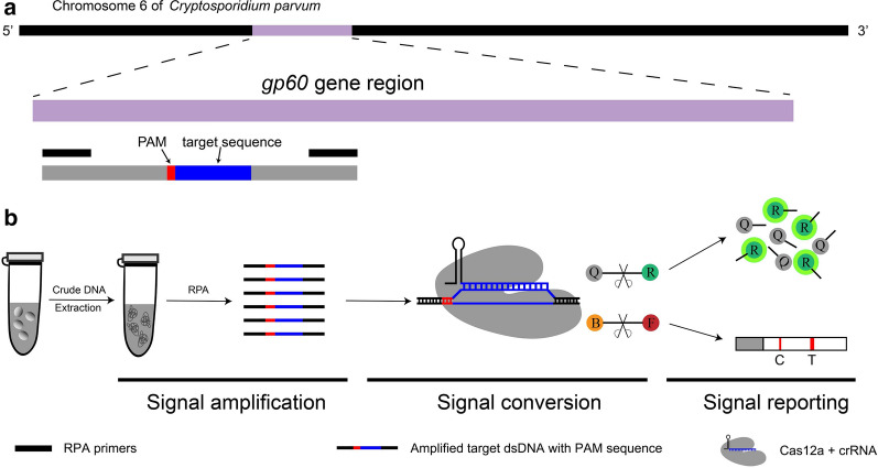
Background: Cryptosporidium parvum is an enteric protozoan parasite with zoonotic importance and can cause cryptosporidiosis in humans as well as domestic and wild animals worldwide. The IId subtype family (SF) is one of the most prevalent subtypes of C. parvum. Some clustered regularly interspaced short palindromic repeats (CRISPR) and CRISPR-associated (Cas) protein systems have been developed to detect nucleic acid with high flexibility, sensitivity and specificity.
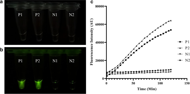
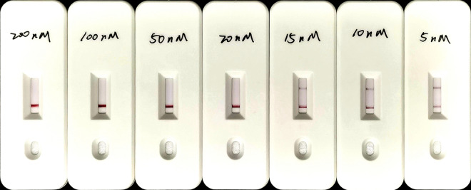
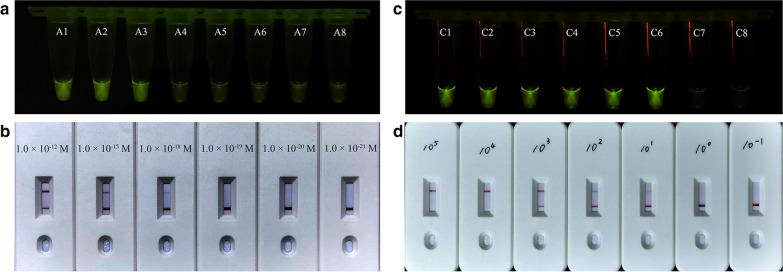
Methods: By integrating recombinase polymerase amplification and the Cas12a/crRNA trans-cleavage system (termed ReCTC), we established end-point diagnostics by observing fluorescence readouts with the naked eye under blue light and on-site diagnostics using a lateral flow strip (LFS) biosensor.
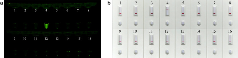
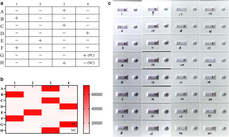
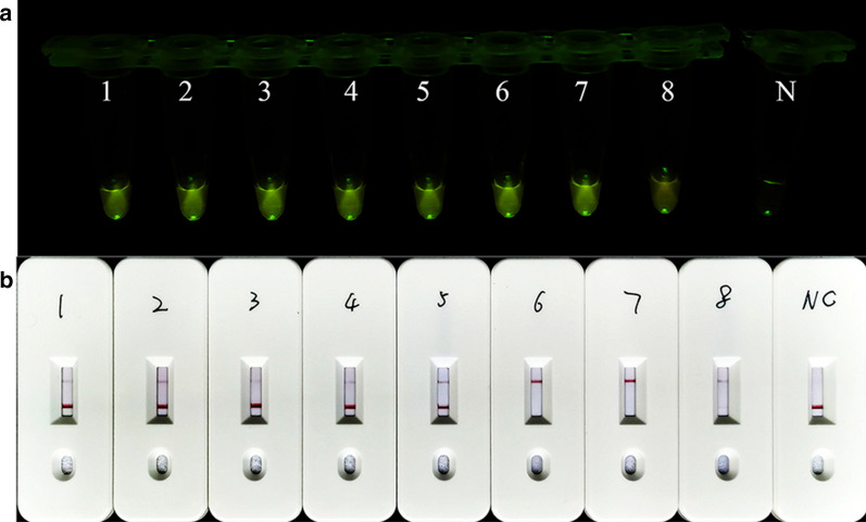
Results: Our ReCTC-based diagnoses can detect as little as a single copy of a cloned C. parvum 60-kDa glycoprotein (GP60) gene, 10 oocysts per gram (OPG), clinical fecal sample without tedious extraction of genomic DNA and have no cross-reactivity with other SFs of C. parvum or other common enteric parasitic protozoa.
Conclusions: This study provided a new strategy for direct identification of the IId SF of C. parvum free of highly trained operators and expensive special equipment.
Keywords: CRISPR/Cas12a; Cryptosporidium parvum; On-site detection; Recombinase polymerase amplification; Visualized detection.
Conflict of interest statement
The authors declare that they have no competing interests.
Figures
Similar articles
-
Prevalence of Cryptosporidium parvum in dairy calves and GP60 subtyping of diarrheic calves in central Argentina.Parasitol Res. 2019 Jul;118(7):2079-2086. doi: 10.1007/s00436-019-06366-y. Epub 2019 Jun 11. Parasitol Res. 2019. PMID: 31187226 Free PMC article.
-
First genetic identification of Cryptosporidium parvum subtype IIaA14G2R1in beef cattle in Brazil.Prev Vet Med. 2015 Oct 1;121(3-4):391-4. doi: 10.1016/j.prevetmed.2015.08.016. Epub 2015 Aug 29. Prev Vet Med. 2015. PMID: 26342791
-
End-point diagnostics of Giardia duodenalis assemblages A and B by combining RPA with CRISPR/Cas12a from human fecal samples.Parasit Vectors. 2024 Nov 12;17(1):463. doi: 10.1186/s13071-024-06559-0. Parasit Vectors. 2024. PMID: 39533301 Free PMC article.
-
Emergence of zoonotic Cryptosporidium parvum in China.Trends Parasitol. 2022 Apr;38(4):335-343. doi: 10.1016/j.pt.2021.12.002. Epub 2021 Dec 28. Trends Parasitol. 2022. PMID: 34972653 Review.
-
Point of care diagnostics for Cryptosporidium: new and emerging technologies.Curr Opin Gastroenterol. 2023 Jan 1;39(1):3-8. doi: 10.1097/MOG.0000000000000895. Epub 2022 Nov 10. Curr Opin Gastroenterol. 2023. PMID: 36504030 Review.
Cited by
-
Development of CRISPR-Mediated Nucleic Acid Detection Technologies and Their Applications in the Livestock Industry.Genes (Basel). 2022 Nov 2;13(11):2007. doi: 10.3390/genes13112007. Genes (Basel). 2022. PMID: 36360244 Free PMC article. Review.
-
Environmental DNA in human and veterinary parasitology - Current applications and future prospects for monitoring and control.Food Waterborne Parasitol. 2022 Nov 13;29:e00183. doi: 10.1016/j.fawpar.2022.e00183. eCollection 2022 Dec. Food Waterborne Parasitol. 2022. PMID: 36419798 Free PMC article.
-
Genetic manipulation for the non-model protozoan Eimeria: Advancements, challenges, and future perspective.iScience. 2025 Feb 17;28(3):112060. doi: 10.1016/j.isci.2025.112060. eCollection 2025 Mar 21. iScience. 2025. PMID: 40109377 Free PMC article. Review.
-
A Multiplex PCR Assay for Simultaneous Detection of Giardia duodenalis, Cryptosporidium parvum, Blastocystis spp. and Enterocytozoon bieneusi in Goats.Vet Sci. 2024 Sep 22;11(9):448. doi: 10.3390/vetsci11090448. Vet Sci. 2024. PMID: 39330827 Free PMC article.
-
CRISPR/Cas12a combined with RPA for detection of T. gondii in mouse whole blood.Parasit Vectors. 2023 Jul 30;16(1):256. doi: 10.1186/s13071-023-05868-0. Parasit Vectors. 2023. PMID: 37518013 Free PMC article.
References
Publication types
MeSH terms
Grants and funding
LinkOut - more resources
Full Text Sources
Other Literature Sources
Medical
