

Infectious Clones Produce SARS-CoV-2 That Causes Severe Pulmonary Disease in Infected K18-Human ACE2 Mice
- PMID: 33879586
- PMCID: PMC8092263
- DOI: 10.1128/mBio.00819-21
Infectious Clones Produce SARS-CoV-2 That Causes Severe Pulmonary Disease in Infected K18-Human ACE2 Mice
Abstract
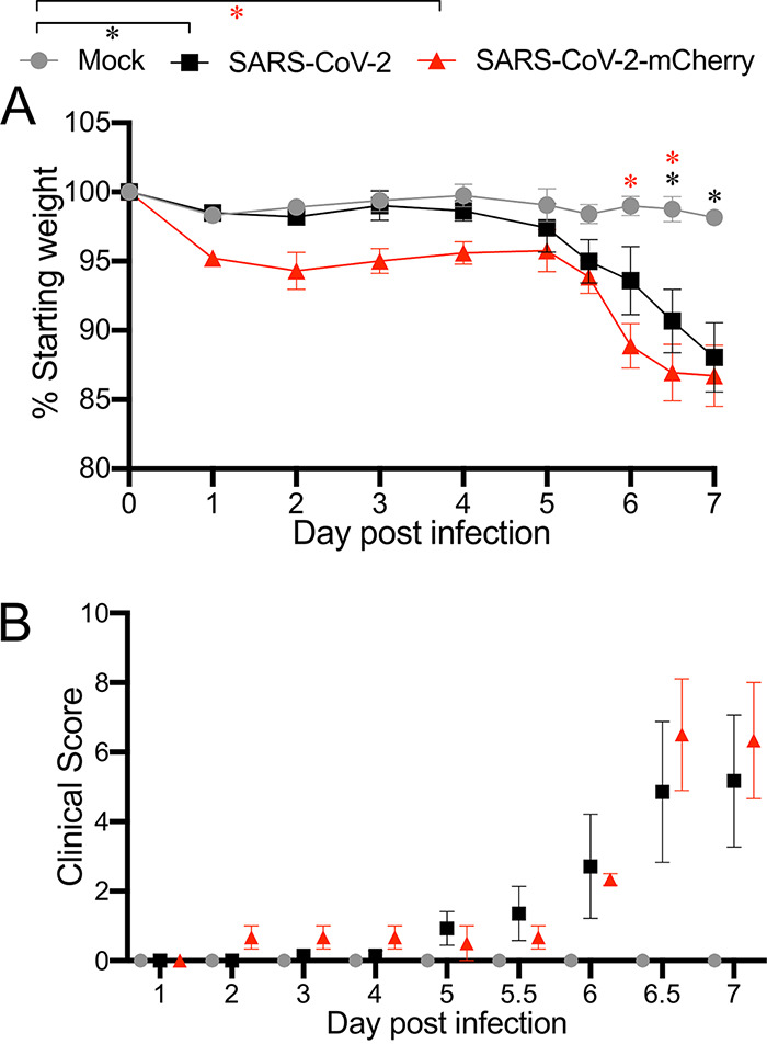
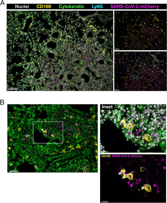
Newly emerged severe acute respiratory syndrome coronavirus 2 (SARS-CoV-2) is the causative agent of the ongoing coronavirus disease 2019 (COVID-19) pandemic, which has caused extensive mortality and morbidity and wreaked havoc on socioeconomic structures. The urgent need to better understand SARS-CoV-2 biology and enable continued development of effective countermeasures is aided by the production of laboratory tools that facilitate SARS-CoV-2 research. We previously created a directly accessible SARS-CoV-2 toolkit containing user-friendly reverse genetic (RG) infectious clones of SARS-CoV-2. Here, using K18-human ACE2 (hACE2) mice, we confirmed the validity of RG-rescued SARS-CoV-2 viruses to reproduce the infection profile, clinical disease, and pathogenesis already established in mice infected with natural SARS-CoV-2 isolates, often patient derived. RG-rescued SARS-CoV-2-infected K18-hACE2 mice developed substantial clinical disease and weight loss by day 6 postinfection. RG-rescued SARS-CoV-2 was recovered from the lungs and brains of infected K18-hACE2 mice, and infection resulted in viral pneumonia with considerable changes in lung pathology, as seen previously with natural SARS-CoV-2 infection. In mice infected with RG-rescued SARS-CoV-2-mCherry, mCherry was detected in areas of lung consolidation and colocalized with clinically relevant SARS-CoV-2-assocated immunopathology. RG-rescued SARS-CoV-2 viruses successfully recapitulated many of the features of severe COVID-19 associated with the K18-hACE2 model of SARS-CoV-2 infection. With utility in vivo, the RG-rescued SARS-CoV-2 viruses will be valuable resources to advance numerous areas of SARS-CoV-2 basic research and COVID-19 vaccine development.IMPORTANCE To develop COVID-19 countermeasures, powerful research tools are essential. We produced a SARS-COV-2 reverse genetic (RG) infectious clone toolkit that will benefit a variety of investigations. In this study, we further prove the toolkit's value by demonstrating the in vivo utility of RG-rescued SARS-CoV-2 isolates. RG-rescued SARS-CoV-2 isolates reproduce disease signs and pathology characteristic of the K18-hACE2 mouse model of severe COVID-19 in infected mice. Having been validated as a model of severe COVID-19 previously using only natural SARS-CoV-2 isolated from patients, this is the first investigation of RG-rescued SARS-CoV-2 viruses in K18-hACE2 mice. The RG-rescued SARS-CoV-2 viruses will facilitate basic understanding of SARS-CoV-2 and the preclinical development of COVID-19 therapeutics.
Keywords: coronavirus; cytokines; infectious clones; lung infection; respiratory viruses.
Copyright © 2021 Liu et al.
Figures


Similar articles
-
SARS-CoV-2 Causes Lung Infection without Severe Disease in Human ACE2 Knock-In Mice.J Virol. 2022 Jan 12;96(1):e0151121. doi: 10.1128/JVI.01511-21. Epub 2021 Oct 20. J Virol. 2022. PMID: 34668780 Free PMC article.
-
A human-ACE2 knock-in mouse model for SARS-CoV-2 infection recapitulates respiratory disorders but avoids neurological disease associated with the transgenic K18-hACE2 model.mBio. 2025 May 14;16(5):e0072025. doi: 10.1128/mbio.00720-25. Epub 2025 Apr 24. mBio. 2025. PMID: 40272151 Free PMC article.
-
The K18-Human ACE2 Transgenic Mouse Model Recapitulates Non-severe and Severe COVID-19 in Response to an Infectious Dose of the SARS-CoV-2 Virus.J Virol. 2022 Jan 12;96(1):e0096421. doi: 10.1128/JVI.00964-21. Epub 2021 Oct 20. J Virol. 2022. PMID: 34668775 Free PMC article.
-
K18- and CAG-hACE2 Transgenic Mouse Models and SARS-CoV-2: Implications for Neurodegeneration Research.Molecules. 2022 Jun 28;27(13):4142. doi: 10.3390/molecules27134142. Molecules. 2022. PMID: 35807384 Free PMC article. Review.
-
Putative mechanism of neurological damage in COVID-19 infection.Acta Neurobiol Exp (Wars). 2021;81(1):69-79. doi: 10.21307/ane-2021-008. Acta Neurobiol Exp (Wars). 2021. PMID: 33949163 Review.
Cited by
-
Human-Immune-System (HIS) humanized mouse model (DRAGA: HLA-A2.HLA-DR4.Rag1KO.IL-2RγcKO.NOD) for COVID-19.Hum Vaccin Immunother. 2022 Nov 30;18(5):2048622. doi: 10.1080/21645515.2022.2048622. Epub 2022 Mar 29. Hum Vaccin Immunother. 2022. PMID: 35348437 Free PMC article.
-
The Delta SARS-CoV-2 Variant of Concern Induces Distinct Pathogenic Patterns of Respiratory Disease in K18-hACE2 Transgenic Mice Compared to the Ancestral Strain from Wuhan.mBio. 2022 Jun 28;13(3):e0068322. doi: 10.1128/mbio.00683-22. Epub 2022 Apr 14. mBio. 2022. PMID: 35420469 Free PMC article.
-
A Rapid Method for Generating Infectious SARS-CoV-2 and Variants Using Mutagenesis and Circular Polymerase Extension Cloning.Microbiol Spectr. 2023 Mar 6;11(2):e0338522. doi: 10.1128/spectrum.03385-22. Online ahead of print. Microbiol Spectr. 2023. PMID: 36877070 Free PMC article.
-
Improved survival of SARS COV-2-infected K18-hACE2 mice treated with adenosine A2AR agonist.Heliyon. 2023 Aug 18;9(8):e19226. doi: 10.1016/j.heliyon.2023.e19226. eCollection 2023 Aug. Heliyon. 2023. PMID: 37664715 Free PMC article.
-
Cytokines and Lipid Mediators of Inflammation in Lungs of SARS-CoV-2 Infected Mice.Front Immunol. 2022 Jun 24;13:893792. doi: 10.3389/fimmu.2022.893792. eCollection 2022. Front Immunol. 2022. PMID: 35812400 Free PMC article.
References
-
- Korber B, Fischer WM, Gnanakaran S, Yoon H, Theiler J, Abfalterer W, Hengartner N, Giorgi EE, Bhattacharya T, Foley B, Hastie KM, Parker MD, Partridge DG, Evans CM, Freeman TM, de Silva TI, Sheffield C-GG, McDanal C, Perez LG, Tang H, Moon-Walker A, Whelan SP, LaBranche CC, Saphire EO, Montefiori DC, Sheffield COVID-19 Genomics Group. 2020. Tracking changes in SARS-CoV-2 spike: evidence that D614G increases infectivity of the COVID-19 virus. Cell 182:812–827.e19. doi:10.1016/j.cell.2020.06.043. - DOI - PMC - PubMed
MeSH terms
Substances
LinkOut - more resources
Full Text Sources
Other Literature Sources
Medical
Molecular Biology Databases
Miscellaneous
