. 2021 Apr 20;12(2):e00264-21.
doi: 10.1128/mBio.00264-21.

Dimer Asymmetry and Light Activation Mechanism in Brucella Blue-Light Sensor Histidine Kinase

Affiliations

Dimer Asymmetry and Light Activation Mechanism in Brucella Blue-Light Sensor Histidine Kinase

Jimena Rinaldi et al. mBio. .

Abstract

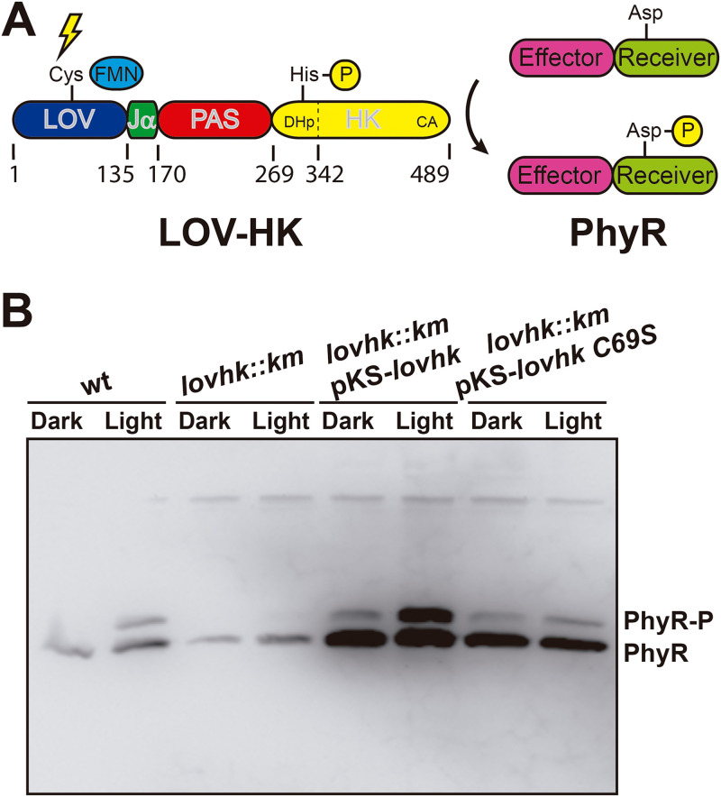
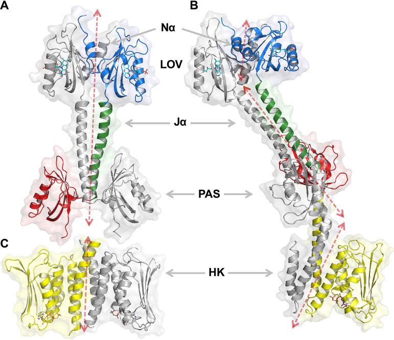
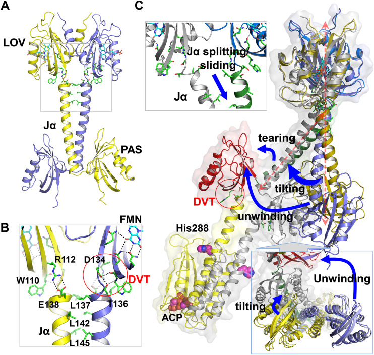
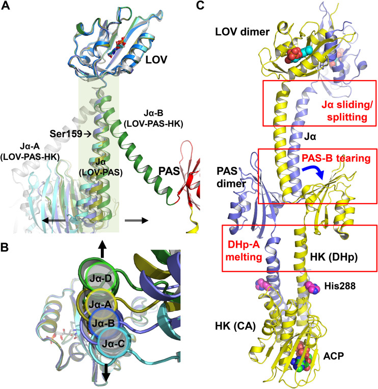
The ability to sense and respond to environmental cues is essential for adaptation and survival in living organisms. In bacteria, this process is accomplished by multidomain sensor histidine kinases that undergo autophosphorylation in response to specific stimuli, thereby triggering downstream signaling cascades. However, the molecular mechanism of allosteric activation is not fully understood in these important sensor proteins. Here, we report the full-length crystal structure of a blue light photoreceptor LOV histidine kinase (LOV-HK) involved in light-dependent virulence modulation in the pathogenic bacterium Brucella abortus Joint analyses of dark and light structures determined in different signaling states have shown that LOV-HK transitions from a symmetric dark structure to a highly asymmetric light state. The initial local and subtle structural signal originated in the chromophore-binding LOV domain alters the dimer asymmetry via a coiled-coil rotary switch and helical bending in the helical spine. These amplified structural changes result in enhanced conformational flexibility and large-scale rearrangements that facilitate the phosphoryl transfer reaction in the HK domain.IMPORTANCE Bacteria employ two-component systems (TCSs) to sense and respond to changes in their surroundings. At the core of the TCS signaling pathway is the multidomain sensor histidine kinase, where the enzymatic activity of its output domain is allosterically controlled by the input signal perceived by the sensor domain. Here, we examine the structures and dynamics of a naturally occurring light-sensitive histidine kinase from the pathogen Brucella abortus in both its full-length and its truncated constructs. Direct comparisons between the structures captured in different signaling states have revealed concerted protein motions in an asymmetric dimer framework in response to light. Findings of this work provide mechanistic insights into modular sensory proteins that share a similar modular architecture.

Keywords: crystallography; dimer asymmetry; light activation mechanism; photoreceptor; sensory histidine kinase.


Figures

FIG 1
FIG 1
In vivo light-gated activation of the two-component system. (A) Schematic representation. The domain architecture of the LOV-HK photoreceptor and the PhyR response regulator is shown, together with the numbering of domain and subdomain boundaries of LOV-HK. (B) Intracellular PhyR∼P levels. The B. abortus 2308 wild-type (wt), the lovhk mutant (lovhk::km), the lovhk mutant complemented with the pKS-lovhk plasmid (lovhk::km pKS-lovhk), and the lovhk mutant complemented with the pKS-lovhk C69S (lovhk::km pKS-lovhk C69S) were grown in dark and light conditions and analyzed by Phos-tag gel electrophoresis and Western blotting with an anti-PhyR antibody. The gel corresponds to one representative experiment of two independent assays.
FIG 2
FIG 2
Crystal structure of LOV-PAS and LOV-PAS-HK. (A) Ribbon diagram of dark-adapted LOV-PAS shows a parallel dimer structure in which the juxtaposed LOV and PAS domains are tethered via two long Jα helices with subtle dimer asymmetry. One subunit is colored according to the domain architecture, while the other is rendered in gray. Ligands are depicted in sticks (see the main text for details). The dashed arrow sketches the trace of the helical spine. (B) Ribbon diagram of light LOV-PAS-HK shows a highly asymmetric dimer with a significant distortion in the helical spine that extends into the HK domain. (C) Ribbon diagram of the isolated HK domain in the inactive state, as published previously (PDB code 5EPV, chains A and B). The location and orientation of this panel give rise, in visual combination with panel A, to an estimate of the structure of the full-length protein in the dark.
FIG 3
FIG 3
Structural comparison between LOV-PAS and LOV-PAS-HK. (A) Coupling between the LOV core and the helical spine in LOV-PAS. (B) A zoom-in view (from the gray box in panel A) highlights the conserved interactions from the FMN binding site to the coiled coil contacts between Jα helices. The FMN isoalloxazine ring is stabilized by a network of H-bonds at the chromophore site. The highly conserved Trp110 residue and the DVT sequence motif are located at the junction between the LOV core and the Jα helix. Two juxtaposed Jα helices are tethered via hydrophobic interactions mediated by Leu137, Leu142, and Leu145 at the dimer interface. (C) Alignment of the LOV-PAS dark structure (in blue/gold) and LOV-PAS-HK light structure according to the LOV dimer reveals a series of structural rearrangements. Blue arrows highlight the structural changes: Jα splitting, tilting of the helical spine, tearing of the PAS domain, and unwinding of the helical spine manifested in a 90° rotation of the PAS dimer. The magenta spheres represent the His288 phosphorylation site and the ATP-analogue molecule bound to the HK domain. The coupling between the PAS domain and the helical spine features a DVT/W motif similar to the LOV domain (red circle).
FIG 4
FIG 4
Dimer asymmetry in the LOV-PAS and LOV-PAS-HK structures. (A) The superposition of the monomer structures from LOV-PAS and LOV-PAS-HK according to the LOV core domain shows dimer asymmetry, which is much less marked in the LOV-PAS dimers than in the LOV-PAS-HK structure (dark green/gray). Subunits A, B, C, and D of LOV-PAS are colored yellow, blue, cyan, and green, respectively. (B) The bottom view of panel A shows small displacements of the Jα helices resulted from dimer asymmetry in LOV-PAS dimers, suggesting that the AB dimer is less asymmetric than the CD dimer. (C) Structural asymmetry in different segments of the LOV-PAS-HK dimer scaffold compared to the LOV-PAS structure.
FIG 5
FIG 5
Light-induced tilting in FMN. (A) In the chromophore site, FMN is stabilized by the conserved residues Asn101, Asn111, and Gln132 with its phosphate group extending out between the Eα and Fα helices. (B) A scatterplot of the top two components from the SVD analysis of 88 simulated annealing omit maps of LOV-PAS near FMN reveals the light-induced signals between 14 dark data sets and 8 light data sets. Each dot corresponds to a map from subunit A (yellow), B (blue), C (cyan), and D (green). The light maps are highlighted with red circles. (C) The decomposed electron density map corresponding to the second component (green, positive density; red, negative density) clearly shows FMN tilting toward Eα upon blue light illumination. The two representative coordinates of dark-adapted (gray) and illuminated (colored) states shown were chosen from the SVD analysis.
FIG 6
FIG 6
Light-induced protein structural changes in LOV-PAS. (A) Alignment of dark structures (in darker shade of yellow/blue) and light structures (in lighter shade of yellow/blue) of LOV-PAS. As FMN tilts outward (blue arrows), the C-terminal ends of the juxtaposed LOV domains partially separate (black arrows). (B) A bottom view of panel A shows that the tethered PAS dimer moves to the same direction (red arrows) in both AB (top) and CD (bottom) dimers. (C) The light-minus-dark difference distance matrix in the LOV domains of AB (left) and CD (right) dimers shows that the distances between the juxtaposed LOV domains increase by 0.5 to 2.0 Å (difference distances are color coded) upon blue light illumination. The LOV domain moves as a rigid body as the intradomain distances remain largely unchanged (small difference distance colored in green). The difference matrices are generated using utilities implemented in dynamiX (36). Both axes of the distance matrix plot represent the residue number of a corresponding LOV domain. In other words, the residue numbers in the x axis from left to right are exactly the same as those in the y axis from top to bottom. For clarity, the LOV domains are labeled along the diagonal of the distance matrix according to their secondary structures (as defined in Fig. S3C) in lieu of the residue numbers on both axes. Cys69 marks the position of the signature Cys residue in the FMN pocket of the LOV domain.
FIG 7
FIG 7
Allosteric activation mechanism proposed for SHK photoreceptors. The dark (left) and light (right) models are represented schematically and colored with the same color code used in Fig. 2. In the box on the right a side view of the PAS dimer in the light state is also shown. We propose a mechanism in which symmetric conformational changes within the LOV domains related to the light signal perception convert into asymmetric transitions related to signal propagation and allosteric activation. Squares, FMN; bars, ADP; triangle, ATP.

References

    1. Buschiazzo A, Trajtenberg F. 2019. Two-component sensing and regulation: how do histidine kinases talk with response regulators at the molecular level? Annu Rev Microbiol 73:507–528. doi:10.1146/annurev-micro-091018-054627. - DOI - PubMed
    1. Jacob-Dubuisson F, Mechaly A, Betton JM, Antoine R. 2018. Structural insights into the signalling mechanisms of two-component systems. Nat Rev Microbiol 16:585–593. doi:10.1038/s41579-018-0055-7. - DOI - PubMed
    1. Gourley CR, Petersen E, Harms J, Splitter G. 2015. Decreased in vivo virulence and altered gene expression by a Brucella melitensis light-sensing histidine kinase mutant. Pathog Dis 73:1–8. doi:10.1111/2049-632X.12209. - DOI - PMC - PubMed
    1. Swartz TE, Tseng TS, Frederickson MA, Paris G, Comerci DJ, Rajashekara G, Kim JG, Mudgett MB, Splitter GA, Ugalde RA, Goldbaum FA, Briggs WR, Bogomolni RA. 2007. Blue-light-activated histidine kinases: two-component sensors in bacteria. Science 317:1090–1093. doi:10.1126/science.1144306. - DOI - PubMed
    1. Alexandre MT, Purcell EB, van Grondelle R, Robert B, Kennis JT, Crosson S. 2010. Electronic and protein structural dynamics of a photosensory histidine kinase. Biochemistry 49:4752–4759. doi:10.1021/bi100527a. - DOI - PMC - PubMed

Publication types

LinkOut - more resources