. 2021 Apr 20;12(5):414.
doi: 10.1038/s41419-021-03692-x.

Drug-resistant cancer cell-derived exosomal EphA2 promotes breast cancer metastasis via the EphA2-Ephrin A1 reverse signaling

Affiliations

Drug-resistant cancer cell-derived exosomal EphA2 promotes breast cancer metastasis via the EphA2-Ephrin A1 reverse signaling

Zicong Gao et al. Cell Death Dis. .

Abstract

Tumor metastasis induced by drug resistance is a major challenge in successful cancer treatment. Nevertheless, the mechanisms underlying the pro-invasive and metastatic ability of drug resistance remain elusive. Exosome-mediated intercellular communications between cancer cells and stromal cells in tumor microenvironment are required for cancer initiation and progression. Recent reports have shown that communications between cancer cells also promote tumor aggression. However, little attention has been regarded on this aspect. Herein, we demonstrated that drug-resistant cell-derived exosomes promoted the invasion of sensitive breast cancer cells. Quantitative proteomic analysis showed that EphA2 was rich in exosomes from drug-resistant cells. Exosomal EphA2 conferred the invasive/metastatic phenotype transfer from drug-resistant cells to sensitive cells. Moreover, exosomal EphA2 activated ERK1/2 signaling through the ligand Ephrin A1-dependent reverse pathway rather than the forward pathway, thereby promoting breast cancer progression. Our findings indicate the key functional role of exosomal EphA2 in the transmission of aggressive phenotype between cancer cells that do not rely on direct cell-cell contact. Our study also suggests that the increase of EphA2 in drug-resistant cell-derived exosomes may be an important mechanism of chemotherapy/drug resistance-induced breast cancer progression.


Conflict of interest statement

The authors declare no competing interests.

Figures
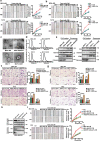
Fig. 1
Fig. 1. Exosomes derived from drug-resistant cells enhance breast cancer cell migration and invasion.
a, b CM from drug-resistant cells significantly enhanced the migratory ability of human breast cancer MDA-MB-468 cells and T47D cells compared with CM from parental cells and fresh medium control as measured by wound healing assay. All experiments were repeated at least three times. ****P < 0.0001. Scale bars: 200 μm. c Transmission electron microscopic image of exosomes derived from drug-resistant cells and its parental cells. Scale bars: 200 nm. d Nanoparticle tracking analysis (NTA) of exosomes derived from drug-resistant cells and its parental cells. e Equal amounts of protein (100 μg) from drug-resistant cells and its parental cells and exosomes were analyzed by Western blotting. Alix, TSG101, and CD81 were used as the positive control of exosomes, and Calnexin was used as the negative control of exosomes. f, g Exosomes derived from drug-resistant cells (DR-Exos) significantly increased the migration and invasion ability of two breast cancer cells compared with exosomes derived from drug-sensitive cells (DS-Exos). For cell migration assay, 5 × 104 cells suspended in 200 μL of serum-free medium were loaded onto the upper chambers. Six hundred microliters of medium with 10% FBS was added into the lower chamber. For cell invasion assay, 1 × 105 cells suspended in 200 μL of serum-free medium were loaded onto the upper chambers coated with Matrigel. The incubation time was 24 h. The statistical results were summarized in the right panel. Data were expressed as mean ± SD. All experiments were repeated at least three times. ****P < 0.0001 and nsP > 0.05 indicate no statistical significance. Scale bars: 200 μm. h Knockdown of Rab27a reduced the amounts of exosomes derived from MDA-MB-468/EPR cells. i Knockdown of Rab27a suppressed the migration-promoting effect of CM derived from drug-resistant cells. All experiments were repeated at least three times. ****P < 0.0001. Scale bars: 200 μm.
Fig. 2
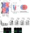
Fig. 2. EphA2 protein is enriched in exosomes derived from drug-resistant cells.
a Heat map of exosomal proteins differentially expressed in MDA-MB-468 and MDA-MB-468/EPR cells. Exosomes were isolated from drug-resistant breast cancer cells and their parental cells, and then a TMT-based quantitative proteomic method was performed to identify differentially expressed proteins in two kinds of exosomes. b Volcano map of exosomal proteins differentially expressed in MDA-MB-468 and MDA-MB-468/EPR cells. Blue and red dots represent the proteins significantly upregulated in exosomes from MDA-MB-468 and MDA-MB-468/EPR cells. c The Venn diagram of different exosomal proteins in MDA-MB-468 cells and MDA-MB-468/EPR cells. d Fifteen proteins were identified by using a strict cutoff (fold change >2.00, score >100). e The expression of EphA2, ABCB1 (encode P-glycoprotein) in exosomes, and cell lysates were analyzed by using Western blotting; β-actin was used as the loading control. f The expression of EphA2 in two drug-resistant breast cancer cells and their parental cells was studied by using immunofluorescence staining. Scale bars: 10 μm.
Fig. 3
Fig. 3. Exosomal EphA2 promotes the migration and invasion of breast cancer cells.
a Western blotting analysis showed that the expression of EphA2 was silenced in drug-resistant cells and exosomes after infection with lentivirus expressing EphA2-specific shRNAs; β-actin was used as the loading control. b, c Exosomes from EphA2-silenced drug-resistant cells disabled the migratory and invasive promoting effect in breast cancer cells. All experiments were repeated at least three times. ****P < 0.0001 and nsP > 0.05 indicate no statistical significance. d Western blotting analysis showed that the expression of EphA2 in HEK-293T cells and exosomes was transfected with control EphA2 vectors. β-actin was used as the loading control. e, f Exosomes from EphA2-expressing HEK-293T cells significantly promoted the migration and invasion ability of breast cancer cells compared with control exosomes. The statistical results were summarized in the right panel. All experiments were repeated at least three times. ****P < 0.0001 and nsP > 0.05 indicate no statistical significance. Scale bars: 200 μm.
Fig. 4
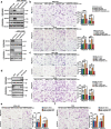
Fig. 4. Exosomal EphA2 promotes migration and invasion of breast cancer cells through the reverse signaling pathway.
a Schematic diagram of the structure of EpA2 mutants. The full-length EphA2 and its three mutants, EphA2-ΔL (deletion of the EphA2 ligand-binding domain), EphA2-ΔS (deletion of the EphA2 SAM domain), and EphA2-S897A (Ser897 mutated to alanine) were cloned into the pCDNA3.1-mCherry vector. b The expression of EphA2 and its mutants was detected in cell lysates and exosomes as measured by Western blotting assay. c The expression of EphA2 and EphA2-S897A was detected in cell lysates and exosomes as measured by Western blotting assay. β-actin was used as the loading control. d, e Flag-tagged Ephrin A1 was co-precipitated with mCherry-tagged EphA2, EphA2-ΔS, and EphA2-S897A but not with EphA2-ΔL mutants. f, g Exosomes carrying EphA2 and EphA2-ΔS, not EphA2-ΔL, promoted the migration and invasion abilities of breast cancer cells. All experiments were repeated at least three times. ****P < 0.0001 and nsP > 0.05 indicate no statistical significance. h Exosomes carrying EphA2-S897A promoted the migration and invasion abilities of breast cancer cells. All experiments were repeated at least three times. ****P < 0.0001 and nsP > 0.05 indicate no statistical significance. Scale bars: 200 μm.
Fig. 5
Fig. 5. Drug-resistant cell-derived exosomal EphA2 promoted the migration and invasion of breast cancer cells by inducing Ephrin A1 reverse signaling.
a Treatment with ALW-II-41-27 suppressed the phosphorylation of EphA2 at the Tyr588 site in drug-resistant breast cancer cells. b Exosomes derived from ALW-II-41-27-treated drug-resistant cells (DR-Exos) exerted migratory promoting effects on sensitive breast cancer cells. The drug-resistant cells were treated with ALW-II-41-27 (500 nM) for 12 h, and then the exosomes were collected and used for further assay. All experiments were repeated at least three times. ****P < 0.0001 and nsP > 0.05 indicate no statistical significance. c Western blotting analysis of the expression of Ephrin A1 in MDA-MB-468 and T47D cells infected with lentivirus expressing control and Ephrin A1-specific shRNAs. d, e Exosomes derived from drug-resistant cells failed to promote the migration and invasion abilities in Ephrin A1 knockdown cells. All experiments were repeated at least three times. **P < 0.01 and nsP > 0.05 indicate no statistical significance. f Exosomes carrying EphA2 and EphA2-ΔS failed to promote the migration of Ephrin A1 knockdown cells. All experiments were repeated at least three times. ****P < 0.0001 and nsP > 0.05 indicate no statistical significance. Scale bars: 200 μm.
Fig. 6
Fig. 6. Drug-resistant cell-derived exosomal EphA2 promotes breast cancer progression through ERK signaling.
a Gene set enrichment analysis (GSEA) showed that the expression of EphA2 significantly correlated with the MAPK signaling pathway based on the TCGA database. b Western blotting analysis of the expression total and phosphorylated Erk1/2, total and phosphorylated Akt, and total and phosphorylated STAT3 in two breast cancer cells treated with exosomes for 24 and 48 h. c Western blotting analysis of the expression of total and phosphorylated Erk1/2 in two breast cancer cells treated with EphA2-rich exosomes derived from HEK-293T cells for 24 h. β-actin was used as the loading control. d Exosomes from EphA2-stable silenced drug-resistant cells failed to induce an elevation of ERK1/2 phosphorylation. e DR-Exos failed to induce an upregulation of phosphorylated ERK1/2 in Ephrin A1 knockdown cells compared with control cells. f Exosomal EphA2 and its mutants EphA2-ΔS could induce upregulation of phosphorylated ERK1/2, whereas exosomal EphA2-ΔL failed to induce ERK1/2 phosphorylation. g Exosomal EphA2 or its mutants cannot induce an increase in ERK1/2 phosphorylation in Ephrin A1 knockdown cells. h PD98059 eliminated the phosphorylation of ERK after incubation with exosomes. i Inhibition of ERK signaling by PD98059 decreased the migration ability of breast cancer cells treated with DR-Exos. All experiments were repeated at least three times. ****P < 0.0001 and nsP > 0.05 indicate no statistical significance. j Exosomes carrying EphA2 and EphA2-ΔS failed to induce an upregulation of phosphorylated ERK1/2 in the presence of PD98095 compared with control cells. k PD98059 blocked the migratory promoting effect of exosomes carrying EphA2 and EphA2-ΔS. All experiments were repeated at least three times. ****P < 0.0001 and nsP > 0.05 indicate no statistical significance. Scale bars: 200 μm.
Fig. 7
Fig. 7. Exosomal EphA2 promotes breast cancer cell metastasis in vivo.
a Schematic diagram of the in vivo experimental design. b Exosomal EphA2 protein was significantly upregulated in plasma from the MDA-MD-468/EPR and MDA-MD-468/EPR-sh control groups compared with the MDA-MD-468 and MDA-MD-468/EPR-EphA2-KD groups. ***P < 0.001 and nsP > 0.05 indicate no statistical significance. c Representative images of subcutaneous tumor formed in mice. d The drug-resistant cell inoculated group showed more metastatic foci on the mice lung surface than that in the drug-sensitive cell inoculated group. Scale bars: 1 cm. e, f H & E staining showed that the number of tumor metastatic foci in the lung tissue was significantly higher in the drug-resistant cell inoculated group than in the drug-sensitive cell inoculated group. Data are shown as mean ± SD. Statistical analysis was performed by one-way ANOVA. ***P < 0.001 and nsP > 0.05 indicate no statistical significance. Scale bars: 200 μm. g The metastatic foci in the lungs of mice were investigated using immunohistochemical (IHC) staining with anti-GFP antibodies. Scale bars: 200 μm. h ELISA assays showed that the concentration of exosomal EphA2 in plasma was collected from healthy donors (n = 20), early-stage breast cancer patients without any systemic treatment (n = 30), and advanced patients received at least one prior line of systemic therapy (n = 30). Data are shown as mean ± SD. Statistical analysis was performed by one-way ANOVA. *P < 0.05, **P < 0.01, ***P < 0.001, and nsP > 0.05 indicate no statistical significance.

Similar articles

Cited by

References

    1. Waks AG, Winer EP. Breast cancer treatment: a review. JAMA. 2019;321:288–300. doi: 10.1001/jama.2018.19323. - DOI - PubMed
    1. Shah AN, Gradishar WJ. Adjuvant anthracyclines in breast cancer: what is their role? Oncologist. 2018;23:1153–1161. doi: 10.1634/theoncologist.2017-0672. - DOI - PMC - PubMed
    1. Jasra S, Anampa J. Anthracycline use for early stage breast cancer in the modern era: a review. Curr. Treat. Options Oncol. 2018;19:30. doi: 10.1007/s11864-018-0547-8. - DOI - PubMed
    1. Hart CD, et al. Challenges in the management of advanced, ER-positive, HER2-negative breast cancer. Nat. Rev. Clin. Oncol. 2015;12:541–552. doi: 10.1038/nrclinonc.2015.99. - DOI - PubMed
    1. Munzone E, Colleoni M. Clinical overview of metronomic chemotherapy in breast cancer. Nat. Rev. Clin. Oncol. 2015;12:631–644. doi: 10.1038/nrclinonc.2015.131. - DOI - PubMed

Publication types

MeSH terms