

Targeting Nanostrategies for Imaging of Atherosclerosis
- PMID: 33880112
- PMCID: PMC8032543
- DOI: 10.1155/2021/6664471
Targeting Nanostrategies for Imaging of Atherosclerosis
Abstract
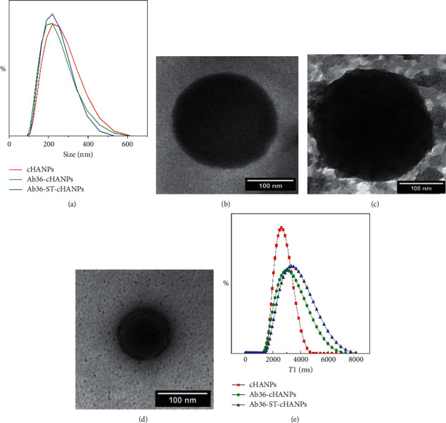
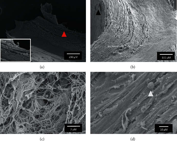
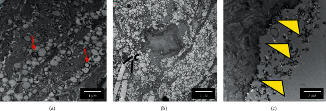
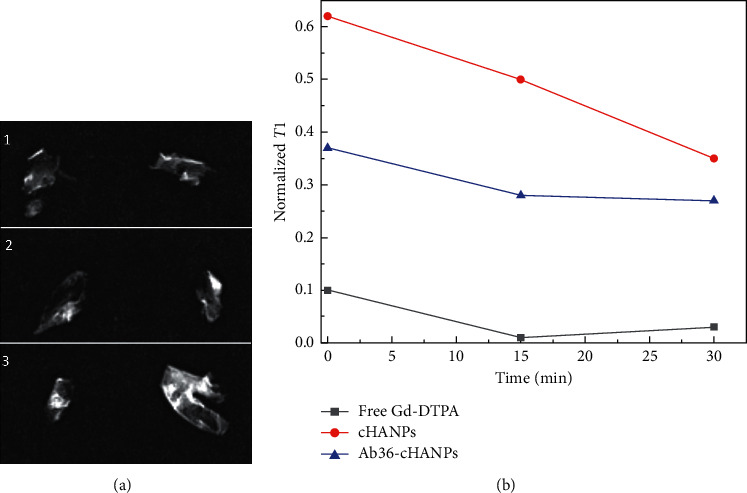
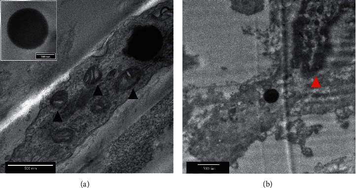
Despite the progress in cardiovascular research, atherosclerosis still represents the main cause of death worldwide. Clinically, the diagnosis of Atherosclerotic Cardiovascular Disease (ASCVD) relies on imaging methodologies including X-ray angiography and computed tomography (CT), which however still fails in the identification of patients at high risk of plaque rupture, the main cause of severe clinical events as stroke and heart attack. Magnetic resonance imaging, which is characterized by very high spatial resolution, could provide a better characterization of atherosclerotic plaque (AP) anatomy and composition, aiding in the identification of "vulnerable" plaques. In this context, hydrogel matrices, which have been demonstrated able to boost relaxometric properties of Gd-based contrast agents (CAs) by the effect of Hydrodenticity, represent a valuable tool towards the precision imaging of ASCVD improving the performance of this class of CAs while reducing systemic toxicity. In particular, hydrogel nanoparticles encapsulating Gd-DTPA can further contribute to providing CA-specific accumulation in the AP by nanoparticle surface decoration triggering an active targeting of the AP with the overall effect of allowing an earlier and more accurate diagnosis. In this work, we tested crosslinked Hyaluronic Acid Nanoparticles (cHANPs) in the complex environment of human atherosclerotic plaque. In addition, the surface of cHANPs was decorated with the antibody anti-CD36 (Ab36-cHANPs) for the active targeting of AP-associated macrophages. Results demonstrate that the Hydrodenticity of cHANPs and Ab36-cHANPs is preserved in this complex system and, preliminarily, that interaction of these probes with the AP is present.
Copyright © 2021 Angela Costagliola di Polidoro et al.
Conflict of interest statement
The authors declare that they have no conflicts of interest regarding the publication of this paper.
Figures
Similar articles
-
A Microfluidic Platform to design crosslinked Hyaluronic Acid Nanoparticles (cHANPs) for enhanced MRI.Sci Rep. 2016 Nov 30;6:37906. doi: 10.1038/srep37906. Sci Rep. 2016. PMID: 27901092 Free PMC article.
-
Commentary on "A Microfluidic Platform to Design Crosslinked Hyaluronic Acid Nanoparticles (cHANPs) for Enhanced MRI".Mol Imaging. 2017 Jan 1;16:1536012117706237. doi: 10.1177/1536012117706237. Mol Imaging. 2017. PMID: 28654388 Free PMC article.
-
Microdistribution of Magnetic Resonance Imaging Contrast Agents in Atherosclerotic Plaques Determined by LA-ICP-MS and SR-μXRF Imaging.Mol Imaging Biol. 2021 Jun;23(3):382-393. doi: 10.1007/s11307-020-01563-z. Epub 2020 Dec 7. Mol Imaging Biol. 2021. PMID: 33289060 Free PMC article.
-
Molecular imaging of atherosclerosis with nanoparticle-based fluorinated MRI contrast agents.Nanomedicine (Lond). 2015;10(11):1817-32. doi: 10.2217/nnm.15.26. Nanomedicine (Lond). 2015. PMID: 26080701 Free PMC article. Review.
-
Magnetic resonance imaging of vulnerable atherosclerotic plaques: current imaging strategies and molecular imaging probes.J Magn Reson Imaging. 2007 Sep;26(3):460-79. doi: 10.1002/jmri.20989. J Magn Reson Imaging. 2007. PMID: 17729343 Review.
Cited by
-
Platelet Membrane Biomimetic Nanoparticles Combined With UTMD to Improve the Stability of Atherosclerotic Plaques.Front Chem. 2022 Mar 8;10:868063. doi: 10.3389/fchem.2022.868063. eCollection 2022. Front Chem. 2022. PMID: 35350774 Free PMC article.
-
Current advances in the imaging of atherosclerotic vulnerable plaque using nanoparticles.Mater Today Bio. 2022 Mar 7;14:100236. doi: 10.1016/j.mtbio.2022.100236. eCollection 2022 Mar. Mater Today Bio. 2022. PMID: 35341094 Free PMC article. Review.
-
Update on the Use of PET/MRI Contrast Agents and Tracers in Brain Oncology: A Systematic Review.Int J Nanomedicine. 2022 Jul 29;17:3343-3359. doi: 10.2147/IJN.S362192. eCollection 2022. Int J Nanomedicine. 2022. PMID: 35937076 Free PMC article.
-
Cardiovascular Tissue Engineering Models for Atherosclerosis Treatment Development.Bioengineering (Basel). 2023 Nov 29;10(12):1373. doi: 10.3390/bioengineering10121373. Bioengineering (Basel). 2023. PMID: 38135964 Free PMC article. Review.
-
Advances in the treatment of atherosclerosis with ligand-modified nanocarriers.Exploration (Beijing). 2023 Dec 7;4(3):20230090. doi: 10.1002/EXP.20230090. eCollection 2024 Jun. Exploration (Beijing). 2023. PMID: 38939861 Free PMC article. Review.
References
Publication types
MeSH terms
Substances
LinkOut - more resources
Full Text Sources
Other Literature Sources
Medical
