

The Clinical Diagnostic Value of Lumbar Intervertebral Disc Herniation Based on MRI Images
- PMID: 33880169
- PMCID: PMC8046570
- DOI: 10.1155/2021/5594920
The Clinical Diagnostic Value of Lumbar Intervertebral Disc Herniation Based on MRI Images
Retraction in
-
Retracted: The Clinical Diagnostic Value of Lumbar Intervertebral Disc Herniation Based on MRI Images.J Healthc Eng. 2023 Oct 11;2023:9792827. doi: 10.1155/2023/9792827. eCollection 2023. J Healthc Eng. 2023. PMID: 37860425 Free PMC article.
Abstract
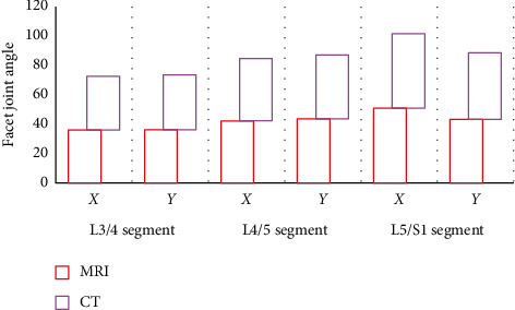
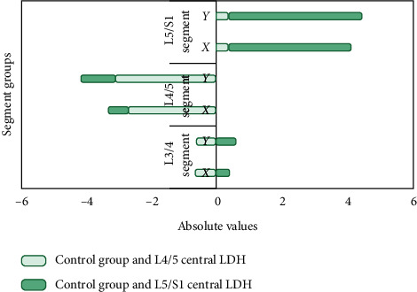
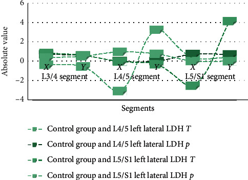
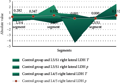
MRI was used to measure the changes in the angle of the facet joints of the lumbar spine and analyze the relationship between it and the herniated lumbar intervertebral disc. Analysis of the causes of lumbar disc herniation from the anatomy and morphology of the spine provides a basis for the early diagnosis and prevention of lumbar disc herniation. There is a certain correlation between the changes shown in MRI imaging of lumbar disc herniation and the TCM syndromes of lumbar intervertebral disc herniation. There is a correlation between the syndromes of lumbar disc herniation and the direct signs of MRI: pathological type, herniated position, and degree of herniation. Indirect signs with MR, nerve root compression and dural sac compression, are related. The MRI examination results can help syndrome differentiation to improve its accuracy to a certain extent. MRI has high sensitivity for the measurement of the angle of the facet joints of the lumbar spine and can be used to study the correlation between the changes of the facet joint angles and the herniated disc. Facet joint asymmetry is closely related to lateral lumbar disc herniation, which may be one of its pathogenesis factors. The herniated intervertebral disc is mostly on the sagittal side of the facet joint, and the facet joint angle on the side of the herniated disc is more sagittal. The asymmetry of the facet joints is not related to the central lumbar disc herniation, and the angle of the facet joints on both sides of the central lumbar disc herniation is partial sagittal.
Copyright © 2021 Kangxing Zheng et al.
Conflict of interest statement
The authors declare that they have no known conflicts of interest or personal relationships that could have appeared to influence the work reported in this paper.
Figures
References
-
- Wu L., Liu L. Analysis of the application value of MRI and CT diagnosis of lumbar disc herniation. Proceedings of Anticancer Research. 2020;4(4):1–10. doi: 10.26689/par.v4i4.1401. - DOI
Publication types
MeSH terms
LinkOut - more resources
Full Text Sources
Other Literature Sources
Medical
