

Intradural extramedullary hemangioblastoma of the thoracic cord: A case report
- PMID: 33880231
- PMCID: PMC8053451
- DOI: 10.25259/SNI_795_2020
Intradural extramedullary hemangioblastoma of the thoracic cord: A case report
Abstract
Background: Spinal hemangioblastomas account for 1-3% of all spinal cord tumors and are mostly intramedullary in location. Here, we report an intradural extramedullary hemangioblastoma of the thoracic spine, occurring in in a patient without von Hippel-Lindau disease.
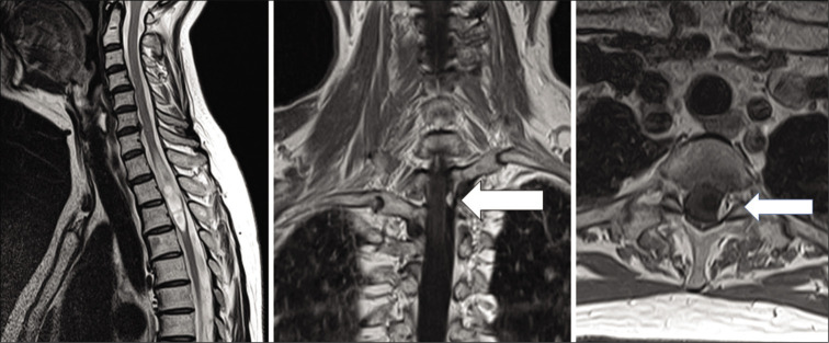
Case description: A 58-year-old female had a 5-year history of progressive left lower extremity weakness. When the MR demonstrated an intradural/extramedullary lesion with a syrinx at the T2-3 level, she successfully underwent gross total tumor excision following which she neurologically improved.
Conclusion: Here, we report a rare case of an intradural/extramedullary thoracic hemangioblastoma successfully excised at the T 2-3 level in a patient without von Hippel-Lindau disease.
Keywords: Cyst; Extramedullary spinal tumor; Hemangioblastoma.
Copyright: © 2021 Surgical Neurology International.
Conflict of interest statement
There are no conflicts of interest.
Figures

Similar articles
-
Intradural Extramedullary Hemangioblastoma of the Cervical Spine: Case Report and Literature Review.Cureus. 2022 May 18;14(5):e25125. doi: 10.7759/cureus.25125. eCollection 2022 May. Cureus. 2022. PMID: 35733499 Free PMC article.
-
Sporadic Intradural Extramedullary Hemangioblastoma of the Cauda Equina: Case Report and Literature Review.World Neurosurg. 2018 Jan;109:436-441. doi: 10.1016/j.wneu.2017.10.104. Epub 2017 Oct 28. World Neurosurg. 2018. PMID: 29107720 Review.
-
Primary Intradural Extramedullary Sporadic Spinal Hemangioblastomas: Case Report and Systematic Review.World Neurosurg. 2021 Aug;152:84-94. doi: 10.1016/j.wneu.2021.05.105. Epub 2021 Jun 1. World Neurosurg. 2021. PMID: 34087464
-
Differentiation of localization of spinal hemangioblastomas based on imaging and pathological findings.Eur Spine J. 2011 Aug;20(8):1377-84. doi: 10.1007/s00586-011-1814-6. Epub 2011 Apr 29. Eur Spine J. 2011. PMID: 21528401 Free PMC article.
-
Intramedullary cervical spinal cord and cerebellar hemangioblastoma: A case report.Surg Neurol Int. 2022 Jul 8;13:294. doi: 10.25259/SNI_525_2022. eCollection 2022. Surg Neurol Int. 2022. PMID: 35855144 Free PMC article.
Cited by
-
Intradural Extramedullary Hemangioblastoma of the Cervical Spine: Case Report and Literature Review.Cureus. 2022 May 18;14(5):e25125. doi: 10.7759/cureus.25125. eCollection 2022 May. Cureus. 2022. PMID: 35733499 Free PMC article.
References
-
- Deng X, Wang K, Wu L, Yang C, Yang T, Zhao L, et al. Intraspinal hemangioblastomas: Analysis of 92 cases in a single institution: Clinical article. J Neurosurg Spine. 2014;21:260–9. - PubMed
-
- Taniguchi S, Ogikubo O, Nakamura T, Yamagishi I, Hayakawa K, Otsuka T, et al. A rare case of extramedullaryintradural hemangioblastoma in the thoracic spine. Spine (Phila Pa 1976) 2009;34:E969–72. - PubMed
-
- Toyoda H, Seki M, Nakamura H, Inoue Y, Yamamoto Y, Takaoka K. Intradural extramedullary hemangioblastoma differentiated by MR images in the cervical spine: A case report and review of the literature. J Spinal Disord Tech. 2004;17:343–7. - PubMed
-
- Yasargil MG, Antic J, Laciga R, de Preux J, Fideler RW, Boone SC. The microsurgical removal of intramedullary spinal hemangioblastomas. Report of twelve cases and a review of the literature. Surg Neurol. 1976;3:141–8. - PubMed
Publication types
LinkOut - more resources
Full Text Sources
Other Literature Sources