

Multiple Artery Aneurysms: Unusual Presentation of IgG4 Vasculopathy
- PMID: 33880242
- PMCID: PMC8053435
- DOI: 10.25259/JCIS_149_2020
Multiple Artery Aneurysms: Unusual Presentation of IgG4 Vasculopathy
Abstract
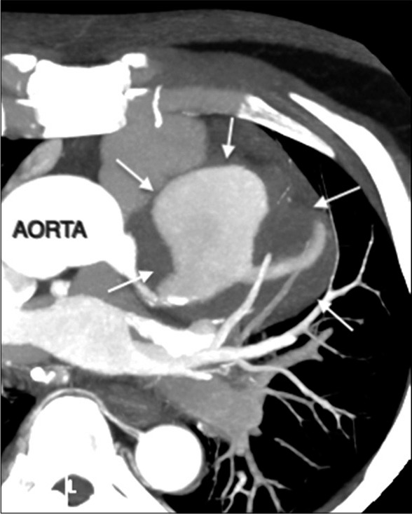
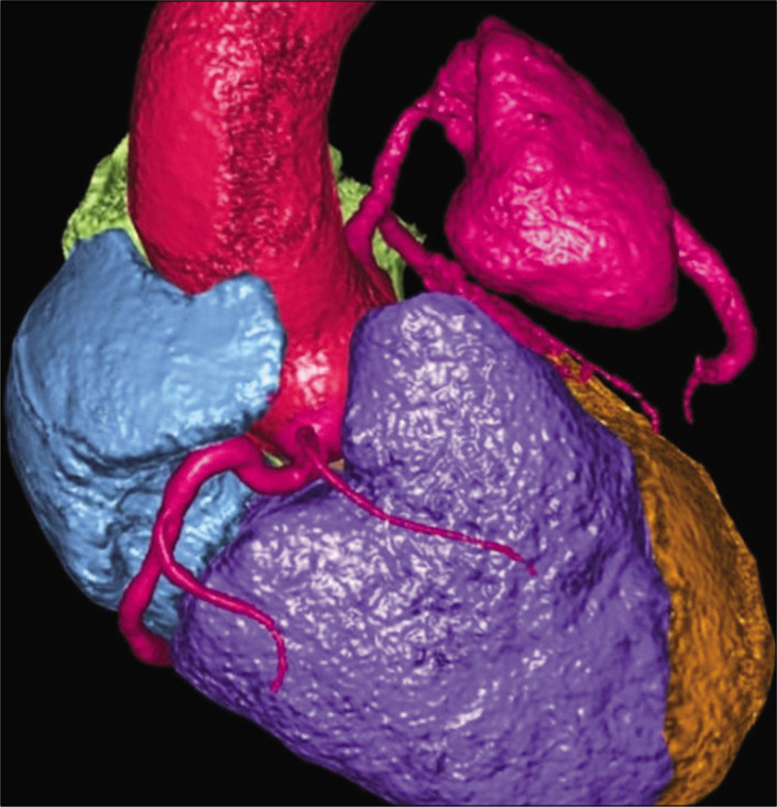
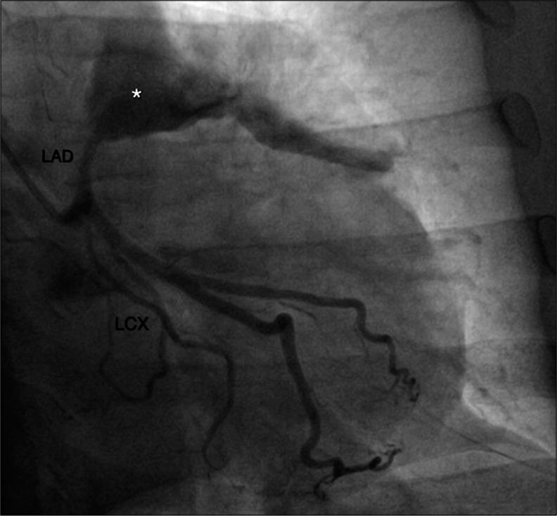
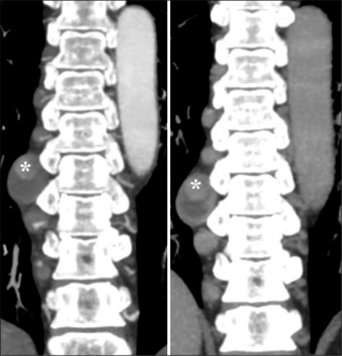
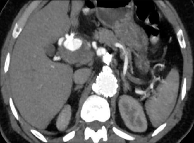
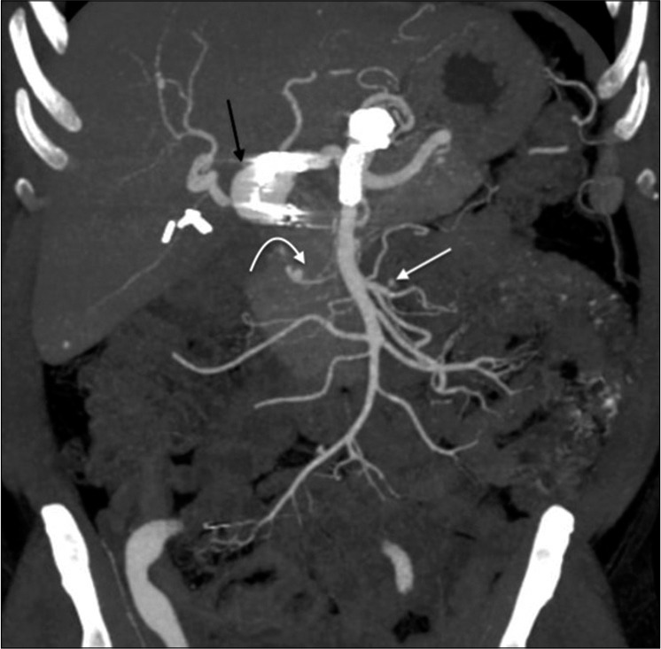
Immunoglobulin G4 (IgG4)-related disease is a chronic systemic disease. It is characterized by inflammatory fibrosis and high serum IgG4 levels. IgG4-positive plasma cells infiltrate target organs in this disease. It may involve the pancreas, biliary tract, lacrimal glands, salivary glands, orbits, thyroid, kidneys, lymph nodes, or retroperitoneum. It may present as vasculitis with involvement of large to medium sized vessels such as the aorta, the common iliac, carotid, and coronary arteries. We present a case of 55-year-old male patient who presented with shortness of breath on exertion and atypical chest pain. On CT angiography, a giant coronary artery aneurysm involving the left anterior descending artery, multiple visceral and intercostal artery aneurysms, and nodular paravertebral soft-tissue thickening secondary to IgG4 vasculopathy.
Keywords: Angiography; Coronary artery aneurysm; Immunoglobulin G4; Vasculitis.
© 2021 Published by Scientific Scholar on behalf of Journal of Clinical Imaging Science.
Conflict of interest statement
There are no conflicts of interest.
Figures
Similar articles
-
An Unusual Presentation of Immunoglobulin G4-Related Disease (IgG4-RD) Causing Subglottic Stenosis.Cureus. 2022 Jun 23;14(6):e26250. doi: 10.7759/cureus.26250. eCollection 2022 Jun. Cureus. 2022. PMID: 35911268 Free PMC article.
-
[Immunoglobulin G4-Related Disease in the Thorax: Imaging Findings and Differential Diagnosis].Taehan Yongsang Uihakhoe Chi. 2021 Jul;82(4):826-837. doi: 10.3348/jksr.2021.0078. Epub 2021 Jul 26. Taehan Yongsang Uihakhoe Chi. 2021. PMID: 36238052 Free PMC article. Review. Korean.
-
A Rare Case of Immunoglobulin G4-Related Disease Presenting With Coronary Artery Pseudotumor And Aneurysm.Eur J Case Rep Intern Med. 2023 Dec 6;11(1):004215. doi: 10.12890/2023_004215. eCollection 2024. Eur J Case Rep Intern Med. 2023. PMID: 38223278 Free PMC article.
-
Immunoglobulin G4-related masses surrounding coronary arteries: a case report.Eur Heart J Case Rep. 2021 Mar 8;5(3):ytab055. doi: 10.1093/ehjcr/ytab055. eCollection 2021 Mar. Eur Heart J Case Rep. 2021. PMID: 34113758 Free PMC article.
-
Immunoglobulin G4-related hepatobiliary disease.Semin Diagn Pathol. 2019 Nov;36(6):423-433. doi: 10.1053/j.semdp.2019.07.007. Epub 2019 Jul 24. Semin Diagn Pathol. 2019. PMID: 31358425 Review.
Cited by
-
Immunoglobulin G4-related hepatic artery aneurysm.J Vasc Surg Cases Innov Tech. 2023 Nov 23;10(1):101377. doi: 10.1016/j.jvscit.2023.101377. eCollection 2024 Feb. J Vasc Surg Cases Innov Tech. 2023. PMID: 38130358 Free PMC article.
-
Reshaping the Concept of Riedel's Thyroiditis into the Larger Frame of IgG4-Related Disease (Spectrum of IgG4-Related Thyroid Disease).Biomedicines. 2023 Jun 11;11(6):1691. doi: 10.3390/biomedicines11061691. Biomedicines. 2023. PMID: 37371786 Free PMC article. Review.
-
Crohn's Disease and Jejunal Artery Aneurysms: A Report of the First Case and a Review of the Literature.Medicina (Kaunas). 2022 Sep 24;58(10):1344. doi: 10.3390/medicina58101344. Medicina (Kaunas). 2022. PMID: 36295505 Free PMC article. Review.
-
Multiple intrahepatic artery aneurysms during the treatment for IgG4-related sclerosing cholangitis: A case report.World J Hepatol. 2024 Dec 27;16(12):1505-1514. doi: 10.4254/wjh.v16.i12.1505. World J Hepatol. 2024. PMID: 39744203 Free PMC article.
-
Coronary Artery Aneurysm or Ectasia as a Form of Coronary Artery Remodeling: Etiology, Pathogenesis, Diagnostics, Complications, and Treatment.Biomedicines. 2024 Sep 2;12(9):1984. doi: 10.3390/biomedicines12091984. Biomedicines. 2024. PMID: 39335497 Free PMC article. Review.
References
-
- Yabusaki S, Oyama-Manabe N, Manabe O, Hirata K, Kato F, Miyamoto N, et al. Characteristics of immunoglobulin G4-related aortitis/periaortitis and periarteritis on fluorodeoxyglucose positron emission tomography/computed tomography co-registered with contrast-enhanced computed tomography. EJNMMI Res. 2017;7:20. doi: 10.1186/s13550-017-0268-1. - DOI - PMC - PubMed
Publication types
LinkOut - more resources
Full Text Sources
Other Literature Sources