

Incidental Malignant Colonic Polyp Detected in a Resected Ischaemic Large Bowel: A Case Report and Literature Review
- PMID: 33880275
- PMCID: PMC8051532
- DOI: 10.7759/cureus.13928
Incidental Malignant Colonic Polyp Detected in a Resected Ischaemic Large Bowel: A Case Report and Literature Review
Abstract
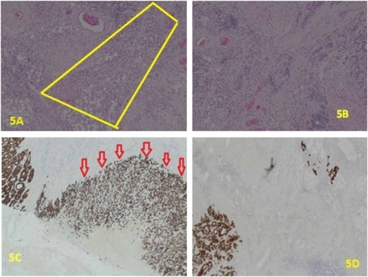
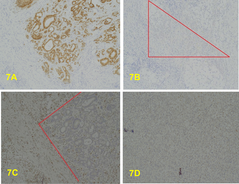
Most patients with bowel cancer are symptomatic at the time of the diagnosis. They may present with a change in bowel habit, bleeding per rectum, abdominal pain, anaemia, weight loss or bowel obstruction. Colonic carcinoma can also be diagnosed incidentally during screening programs. Moreover, it may be incidentally detected in CT scans being performed for other indications or encountered during surgery for other causes. Some patients with colonic bowel ischaemia have associated large bowel cancer, where the ischaemic segment is usually proximal to the tumour and not necessarily associated with bowel obstruction. We are presenting a rare case of incidental malignant colonic polyp detected in a resected ischaemic large bowel in an 88-year-old gentleman. This was a very small tumour that was not visible macroscopically or detectable by imaging. Pathological examination of non-tumour colorectal resection specimens, as in this case, should include careful macroscopic examination and sequential block selection along the length of the colon, and where there is diffuse mucosal abnormality, block selection at 100mm interval is also advised. Attention to and block selection from any suspicious-looking area is warranted in all cases of non-tumour colorectal resections if such microscopic-sized malignancies of the type seen in our patient are to be picked up.
Keywords: adenocarcinoma; colonic cancer; colorectal cancer; laparotomy; lynch syndrome; malignant colonic polyp.
Copyright © 2021, Idaewor et al.
Conflict of interest statement
The authors have declared that no competing interests exist.
Figures


Similar articles
-
[Intestinal occlusion due to a colonic lipoma. Apropos 2 cases].Minerva Chir. 1993 Sep 30;48(18):1035-9. Minerva Chir. 1993. PMID: 8290148 Italian.
-
Dual Bowel Obstruction: A Rare Case of Gallstone Ileus and Colonic Adenocarcinoma.Cureus. 2022 Jan 18;14(1):e21379. doi: 10.7759/cureus.21379. eCollection 2022 Jan. Cureus. 2022. PMID: 35198291 Free PMC article.
-
Early Discovery Of Small Bowel Adenocarcinoma In a Patient Admitted For 4 Acute Intestinal Intussusception.Ann Med Surg (Lond). 2022 Sep 22;82:104776. doi: 10.1016/j.amsu.2022.104776. eCollection 2022 Oct. Ann Med Surg (Lond). 2022. PMID: 36268363 Free PMC article.
-
Hamartomatous polyps - a clinical and molecular genetic study.Dan Med J. 2016 Aug;63(8):B5280. Dan Med J. 2016. PMID: 27477802 Review.
-
Colonic mucosubmucosal elongated polyp: a clinicopathologic study of 13 cases and review of the literature.Am J Surg Pathol. 2011 Dec;35(12):1818-22. doi: 10.1097/PAS.0b013e31822c0688. Am J Surg Pathol. 2011. PMID: 21989340 Review.
References
Publication types
LinkOut - more resources
Full Text Sources
Other Literature Sources