. 2021 Apr 8;2(2):100026.
doi: 10.1016/j.xhgg.2021.100026. Epub 2021 Jan 30.

Genotype and defects in microtubule-based motility correlate with clinical severity in KIF1A-associated neurological disorder

Affiliations

Genotype and defects in microtubule-based motility correlate with clinical severity in KIF1A-associated neurological disorder

Lia Boyle et al. HGG Adv. .

Abstract

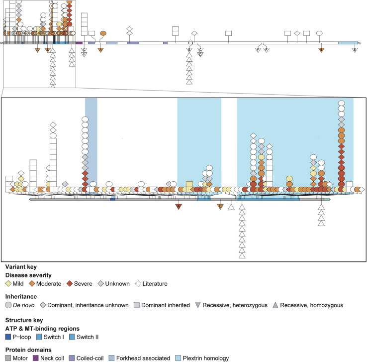
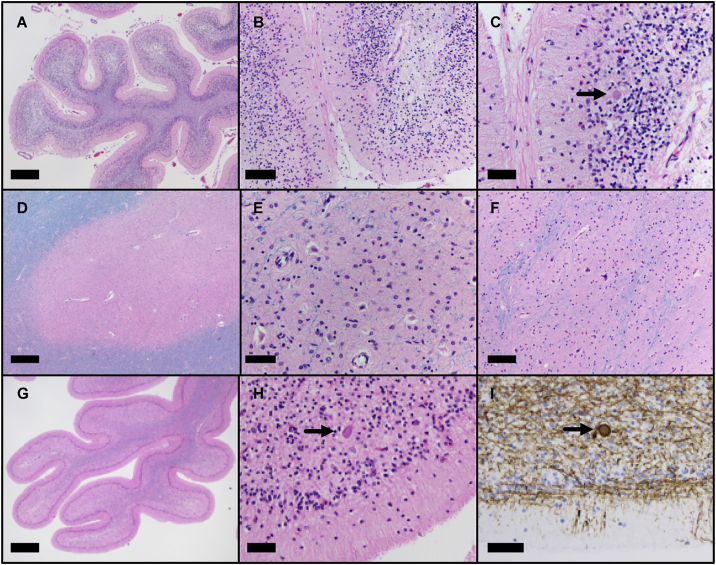
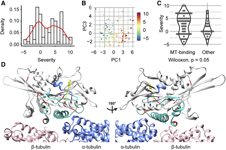
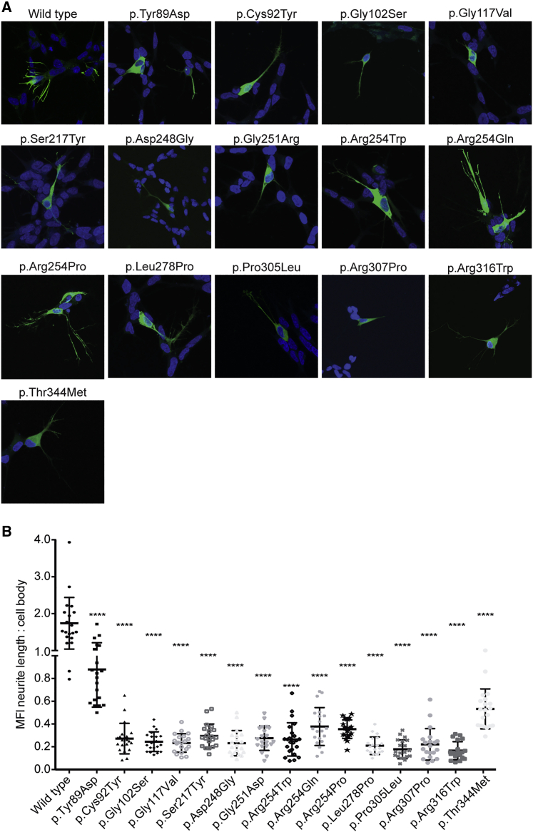
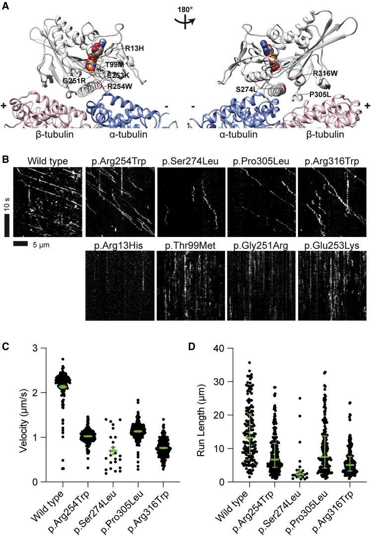
KIF1A-associated neurological disorder (KAND) encompasses a group of rare neurodegenerative conditions caused by variants in KIF1A,a gene that encodes an anterograde neuronal microtubule (MT) motor protein. Here we characterize the natural history of KAND in 117 individuals using a combination of caregiver or self-reported medical history, a standardized measure of adaptive behavior, clinical records, and neuropathology. We developed a heuristic severity score using a weighted sum of common symptoms to assess disease severity. Focusing on 100 individuals, we compared the average clinical severity score for each variant with in silico predictions of deleteriousness and location in the protein. We found increased severity is strongly associated with variants occurring in protein regions involved with ATP and MT binding: the P loop, switch I, and switch II. For a subset of variants, we generated recombinant proteins, which we used to assess transport in vivo by assessing neurite tip accumulation and to assess MT binding, motor velocity, and processivity using total internal reflection fluorescence microscopy. We find all modeled variants result in defects in protein transport, and we describe three classes of protein dysfunction: reduced MT binding, reduced velocity and processivity, and increased non-motile rigor MT binding. The rigor phenotype is consistently associated with the most severe clinical phenotype, while reduced MT binding is associated with milder clinical phenotypes. Our findings suggest the clinical phenotypic heterogeneity in KAND likely reflects and parallels diverse molecular phenotypes. We propose a different way to describe KAND subtypes to better capture the breadth of disease severity.


Conflict of interest statement

Declaration of interests The authors declare no competing interests.

Figures

Figure 1
Figure 1
KIF1A variants by severity Shapes representing 251 affected individuals are shown below, including 114 individuals from our cohort and 137 individuals previously reported in the literature. Disease severity is shown for 98 of these individuals from our cohort. Four affected individuals are not incorporated, as their variants are not mappable below; this includes 3 variants affecting splicing (2 in our cohort, 1 in the literature) and one larger deletion from the literature. On top is the full-length protein, and on the bottom is a zoom in of the motor domain. Each icon represents a unique individual. The shape indicates inheritance and color indicates disease severity. In both the full-length protein and the motor domain, monoallelic variants are shown above and biallelic variants are shown below. Compound heterozygous individuals are represented by two separate triangles, one at each variant; those triangles have letters to indicate variant pairing. ATP and MT binding regions in the motor domain are indicated by color. Splicing information is only indicated for the alternatively spliced exon 25b.
Figure 2
Figure 2
Neuropathological changes in two individuals with c.296C>T (p.Thr99Met) KIF1A variant Microscopic findings from a 13-year-old female (A–F) and a 6-year-old female (G–I). Microscopic examination of H&E/Luxol fast blue-stained tissue sections reveal severely atrophic cerebellar folia (A and G) with thinned molecular layer, depletion of Purkinje cells, depletion of internal granule cells, Bergmann gliosis, and pallor of the white matter (B, C, and H). These changes are most severe in the superior aspect of the cerebellar vermis but are present in varying degrees throughout the cerebellum. Rare axonal spheroids are present (C and H, arrow) highlighted by neurofilament immunostain (I). The dentate nucleus (D and E) as well as the inferior olivary nucleus of the medulla (F) show marked loss of neurons and severe reactive gliosis. Residual neurons in these areas are hypereosinophilic with pyknotic nuclei. Scale bars: 1 mm (20× magnification) (A, D, and G), 200 μm (100× magnification) (B and F), 100 μm (200× magnification) (C, E, H, and I).
Figure 3
Figure 3
Clinical severity of 100 KAND individuals (A) Distribution of heuristic severity score. (B) Scatterplot of principle components 1 and 3 color-coded by medical history-derived severity scores. (C) Violin plot of medical history-derived severity scores for individuals with variants in or outside of ATP or MT binding regions. (D) Structure of the MT-bound KIF1A motor domain with severe residues and functional regions involved in ATP and MT binding highlighted (PDB: 4UXP, H. sapiens KIF1A CryoEM structure in the presence of AMP-PNP). Light gray: KIF1A; blue: α-tubulin; pink: β-tubulin; yellow: AMP-PNP; red: residues mutated in most severe variants (p.Thr99, p.Glu148, p.Leu157, p.Val186, p.Ser214, p.Ser215, p.Gly251, p.Glu253, p.Arg254, p.Tyr306, p.Arg307, p.Arg316). The P loop, switch I, and switch II are shown in light blue, cyan, and teal, respectively.
Figure 4
Figure 4
Neurite tip accumulation in differentiated SH-SY5Y cells for KIF1A variants seen in KAND individuals (A) Rat KIF1A (1–393) tagged with 3xmCit tag (green) accumulated in the distal tips of differentiated SH-SY5Y cells in case of WT but was localized closer to the cell body in cells transfected with KIF1A constructs containing individual variants. (B) A significant reduction in the mean florescence intensity (MFI) along the neurite length versus the cell body was observed in the 15 individual variants tested. Data were analyzed from three independent experiments using a Mann-Whitney test (n = 20 for each experiment) and are plotted as mean ± SD. ∗∗∗∗p < 0.0001 versus WT.
Figure 5
Figure 5
Sites of KIF1A mutations and motility analyses of dimeric KIF1A motors (A) Structure of the MT-bound KIF1A motor domain with the sites of mutated residues used for TIRF microscopy. Light gray, KIF1A; blue, α-tubulin; pink, β-tubulin. Van der Walls sphere represents AMP-PNP; red residues represent the mutation site (PDB: 4UXP, H. sapiens KIF1A CryoEM structure in the presence of AMP-PNP). p.Arg13His, p.Thr99Met, p.Gly251Arg, p.Glu253Lys, and p.Arg254Trp are at or close to the ATP binding site of KIF1A (left). p.Ser274Leu, p.Pro305Leu, and p.Arg316Trp are close to the β-tubulin binding interface (right). (B) Kymographs of KIF1A WT and mobile (top) and non-motile (bottom) motors. Diagonal lines in the kymograph represent KIF1A molecules that are moving over time. The depicted scale bars are the same for all kymographs shown in this figure. (C) Velocities of KIF1A WT and the mobile motors. The green bars indicate the mean with SEM. Compared to WT, all the aberrant motors have reduced velocities. The WT has an average velocity of 2.1 ± 0.03 μm/s (mean ± SEM) (n = 162), while p.Arg254Trp, p.Ser274Leu, p.Pro305Leu, and p.Arg316Trp move at 1.0 ± 0.01 μm/s (n = 214), 0.7 ± 0.08 μm/s (n = 24), 1.1 ± 0.01 μm/s (n = 181), and 0.8 ± 0.01 μm/s (n = 210), respectively. (D) Run lengths of KIF1A WT and aberrant motors. The green bars indicate the median with quartile. All the aberrant motors have reduced run lengths. The WT’s processivity has a median value of 13.2 (8.3, 20.4) μm, while p.Arg254Trp, p.Ser274Leu, p.Pro305Leu, and p.Arg316Trp have run lengths of 6.7 (4.4, 11.6) μm, 2.3 (1.5, 3.5) μm, 7.5 (4.1, 13.8) μm, and 5.1 (3.2, 7.7) μm, respectively.

Similar articles

Cited by

References

    1. Alfadhel M., Yong S.L., Lillquist Y., Langlois S. Precocious puberty in two girls with PEHO syndrome: a clinical feature not previously described. J. Child Neurol. 2011;26:851–857. - PubMed
    1. Rivière J.-B., Ramalingam S., Lavastre V., Shekarabi M., Holbert S., Lafontaine J., Srour M., Merner N., Rochefort D., Hince P., et al. KIF1A, an axonal transporter of synaptic vesicles, is mutated in hereditary sensory and autonomic neuropathy type 2. Am. J. Hum. Genet. 2011;89:219–230. - PMC - PubMed
    1. Erlich Y., Edvardson S., Hodges E., Zenvirt S., Thekkat P., Shaag A., Dor T., Hannon G.J., Elpeleg O. Exome sequencing and disease-network analysis of a single family implicate a mutation in KIF1A in hereditary spastic paraparesis. Genome Res. 2011;21:658–664. - PMC - PubMed
    1. Hamdan F.F., Gauthier J., Araki Y., Lin D.T., Yoshizawa Y., Higashi K., Park A.R., Spiegelman D., Dobrzeniecka S., Piton A., et al. S2D Group Excess of de novo deleterious mutations in genes associated with glutamatergic systems in nonsyndromic intellectual disability. Am. J. Hum. Genet. 2011;88:306–316. - PMC - PubMed
    1. Klebe S., Lossos A., Azzedine H., Mundwiller E., Sheffer R., Gaussen M., Marelli C., Nawara M., Carpentier W., Meyer V., et al. KIF1A missense mutations in SPG30, an autosomal recessive spastic paraplegia: distinct phenotypes according to the nature of the mutations. Eur. J. Hum. Genet. 2012;20:645–649. - PMC - PubMed

LinkOut - more resources