[Preprint]. 2021 Apr 14:2021.04.14.439844.
doi: 10.1101/2021.04.14.439844.

The SARS-CoV-2 mRNA-1273 vaccine elicits more RBD-focused neutralization, but with broader antibody binding within the RBD

Affiliations

The SARS-CoV-2 mRNA-1273 vaccine elicits more RBD-focused neutralization, but with broader antibody binding within the RBD

Allison J Greaney et al. bioRxiv. .

Update in

Abstract

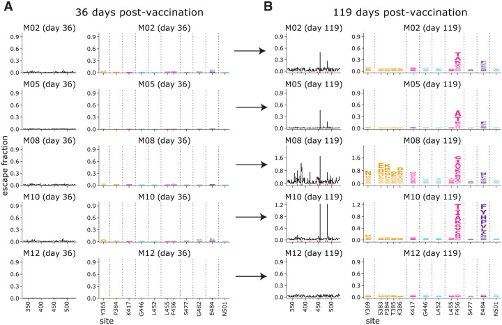
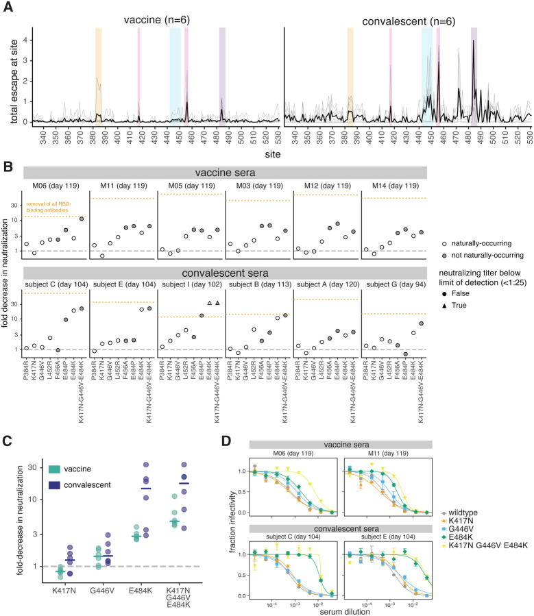
The emergence of SARS-CoV-2 variants with mutations in key antibody epitopes has raised concerns that antigenic evolution will erode immunity. The susceptibility of immunity to viral evolution is shaped in part by the breadth of epitopes targeted. Here we compare the specificity of antibodies elicited by the mRNA-1273 vaccine versus natural infection. The neutralizing activity of vaccine-elicited antibodies is even more focused on the spike receptor-binding domain (RBD) than for infection-elicited antibodies. However, within the RBD, binding of vaccine-elicited antibodies is more broadly distributed across epitopes than for infection-elicited antibodies. This greater binding breadth means single RBD mutations have less impact on neutralization by vaccine sera than convalescent sera. Therefore, antibody immunity acquired by different means may have differing susceptibility to erosion by viral evolution.

One sentence summary: Deep mutational scanning shows the mRNA-1273 RBD-binding antibody response is less affected by single viral mutations than the infection response.


Conflict of interest statement

Competing interests:

HYC is a consultant for Merck, Pfizer, Ellume, and Bill and Melinda Gates Foundation and has received support from Cepheid and Sanofi-Pasteur. The other authors declare no competing interests.

Figures

Fig. 1.
Fig. 1.. RBD-binding antibodies are responsible for most neutralizing activity of mRNA-1273 vaccine-elicited sera.
(A) Binding of serum antibodies to SARS-CoV-2 RBD, as measured by ELISA area-under-the-curve (AUC), for vaccine-elicited sera and convalescent plasmas before and after depletion of RBD-binding antibodies. The dashed pink line indicates binding of pre-pandemic sera. (B) Binding of serum antibodies to the full spike ectodomain. The y-axis scale units in (A) and (B) are not comparable between vaccine and convalescent samples due to different dilution factors (beginning at 1:500 for vaccine sera and 1:100 for convalescent plasmas). (C) Neutralization titer (reciprocal IC50) of vaccine-elicited sera and convalescent plasmas before and after depletion of RBD-binding antibodies. The limit of detection is shown as a dashed horizontal pink line. (D) Percent of neutralizing activity of vaccine-elicited sera and convalescent plasmas due to RBD-binding antibodies. P-values are from a log-rank test accounting for censoring. n=17 for each time point for convalescent plasmas, and n=14 for each time point for vaccine sera. All measurements of convalescent plasma binding and neutralization were previously reported in (15).
Fig. 2.
Fig. 2.. Complete maps of RBD mutations that reduce binding by serum collected 119 days post-vaccination with the 250 μg dose.
(A) The epitopes of four major classes (24) of RBD-binding antibodies are colored on the RBD surface (PDB 6M0J). ACE2 is shown as a gray cartoon. (B) Number of sera that fell into each of the four major categories of binding-escape maps as categorized by subjective visual inspection. (C) Escape maps for six representative sera. The line plots at left indicate the sum of effects of all mutations at each RBD site on serum antibody binding, with larger values indicating more escape. The logo plots at right show key sites (highlighted in purple on the line plot x-axes). The height of each letter is that mutation’s escape fraction; larger letters indicate a greater reduction in binding. Escape fractions are not strictly comparable between samples due to the use of sample-specific FACS selection gates—therefore, for each sample, the y-axis is scaled independently (see Methods). RBD sites are colored by epitope as in (A). The escape fractions were well-correlated between independent libraries, and we report the average of duplicate measurements throughout (Table S3, Fig. S4). Escape maps for all 14 sera collected at day 119 from individuals who received the 250 μg dose are shown in Fig. S5. Comparable escape maps for eight individuals who received the 100 μg dose are in Fig. S6. Interactive versions of logo plots and structural visualizations are at https://jbloomlab.github.io/SARS-CoV-2-RBD_MAP_Moderna/.
Fig. 3.
Fig. 3.. Comparison of escape maps for sera collected at day 36 and day 119 post-vaccination shows that the RBD-binding response focuses over time.
Escape maps for sera at day 36 (A) and day 119 (B) from 5 individuals who received the 250 μg vaccine dose. The day 36 maps are all relatively flat, indicating no RBD mutation has a large effect on serum antibody binding. By day 119, the maps are often more focused on sites 456 and 484. The y-axis is scaled separately for each serum (see Methods). Interactive versions are at https://jbloomlab.github.io/SARS-CoV-2-RBD_MAP_Moderna/.
Fig. 4.
Fig. 4.. The binding of vaccine-elicited polyclonal antibodies is more broadly distributed across the RBD than for infection-elicited antibodies.
(A) Escape from RBD-binding antibodies at each site in the RBD for vaccine sera or convalescent plasmas collected at early or late time points. Thin gray lines show individual serum or plasma samples, and the thick black line shows the mean (number of samples is indicated in the plot titles). Key sites within the epitopes of each major RBD antibody class are highlighted with the colors defined in Fig. 2A and in panel (B). (B) Relationships among escape maps of vaccine sera, convalescent plasmas, and monoclonal antibodies visualized with a multidimensional scaling projection. Vaccine sera include both doses and time points. Convalescent plasmas include all time points. (C) Binding escape at each site mapped onto the RBD surface after averaging across all sera/plasmas in each group. The RBD surface coloring is scaled from white to red, with white indicating no escape, and red indicating the site with the greatest escape. The color scaling spans the full range of white to red for each serum/plasma group, so the quantitative scale is not comparable across groups. Escape maps for monoclonal antibodies previously described in (, , –27), and convalescent plasmas in (15, 16).
Fig. 5.
Fig. 5.. Effects of RBD mutations on neutralization by day 100–150 sera from vaccinated and convalescent individuals.
(A) Total binding escape at each RBD site is shown for the six vaccine and convalescent samples tested in neutralization assays. The thin gray lines show individual samples, and the dark black line shows the mean. Key sites within each epitope are highlighted using the same color scheme as in Fig. 2A. (B) Neutralization of G614 spike-pseudotyped lentiviral particles with the indicated RBD mutations, shown as the fold-increase in IC50 compared to G614 spike with no additional mutations. Mutations that have been observed in human SARS-CoV-2 isolates are colored in white, and non-naturally-occurring mutations in gray. The orange dashed line represents the effect of depleting all RBD-binding antibodies, as in Fig. 1. (C) The fold decrease in neutralization titer caused by individual mutations in each of the three major neutralizing epitopes of the RBD (K417 in the class 1 epitope, E484K in the class 2 epitope, and G446V in the class 3 epitope), as well as the combination of all three mutations. Horizontal lines represent the median. In (B) and (C), the dashed gray line indicates no change in neutralization relative to unmutated spike. (D) Representative neutralization curves from two vaccine and two convalescent samples against the triple mutant and its composite single mutations. IC50s and pseudovirus entry titers are shown in Fig. S8 and all neutralization curves are in Fig. S9.

References

    1. Schmidt O. W., Allan I. D., Cooney M. K., Foy H. M., Fox J. P., Rises in titers of antibody to human coronaviruses OC43 and 229E in Seattle families during 1975–1979. Am. J. Epidemiol. 123, 862–868 (1986). - PMC - PubMed
    1. Eguia R. T., Crawford K. H. D., Stevens-Ayers T., Kelnhofer-Millevolte L., Greninger A. L., Englund J. A., Boeckh M. J., Bloom J. D., A human coronavirus evolves antigenically to escape antibody immunity. PLoS Pathog. 17, e1009453 (2021). - PMC - PubMed
    1. Cele S., Gazy I., Jackson L., Hwa S.-H., Tegally H., Lustig G., Giandhari J., Pillay S., Wilkinson E., Naidoo Y., Karim F., Ganga Y., Khan K., Bernstein M., Balazs A. B., Gosnell B. I., Hanekom W., Moosa M.-Y. S., NGS-SA, COMMIT-KZN Team, Lessells R. J., de Oliveira T., Sigal A., Escape of SARS-CoV-2 501Y.V2 from neutralization by convalescent plasma. Nature, 2021.01.26.21250224 (2021). - PMC - PubMed
    1. Garcia-Beltran W. F., Lam E. C., St Denis K., Nitido A. D., Garcia Z. H., Hauser B. M., Feldman J., Pavlovic M. N., Gregory D. J., Poznansky M. C., Sigal A., Schmidt A. G., Iafrate A. J., Naranbhai V., Balazs A. B., Multiple SARS-CoV-2 variants escape neutralization by vaccine-induced humoral immunity. Cell (2021), doi: 10.1016/j.cell.2021.03.013 - DOI - PMC - PubMed
    1. Wang Z., Schmidt F., Weisblum Y., Muecksch F., Barnes C. O., Finkin S., Schaefer-Babajew D., Cipolla M., Gaebler C., Lieberman J. A., Oliveira T. Y., Yang Z., Abernathy M. E., Huey-Tubman K. E., Hurley A., Turroja M., West K. A., Gordon K., Millard K. G., Ramos V., Silva J. D., Xu J., Colbert R. A., Patel R., Dizon J., Unson-O’Brien C., Shimeliovich I., Gazumyan A., Caskey M., Bjorkman P. J., Casellas R., Hatziioannou T., Bieniasz P. D., Nussenzweig M. C., mRNA vaccine-elicited antibodies to SARS-CoV-2 and circulating variants. Nature, 2021.01.15.426911 (2021). - PMC - PubMed

Publication types