. 2021 Jul 28;144(6):1833-1852.
doi: 10.1093/brain/awab062.

A delta-secretase-truncated APP fragment activates CEBPB, mediating Alzheimer's disease pathologies

Affiliations

A delta-secretase-truncated APP fragment activates CEBPB, mediating Alzheimer's disease pathologies

Yinan Yao et al. Brain. .

Abstract

Amyloid-β precursor protein (APP) is sequentially cleaved by secretases and generates amyloid-β, the major components in senile plaques in Alzheimer's disease. APP is upregulated in human Alzheimer's disease brains. However, the molecular mechanism of how APP contributes to Alzheimer's disease pathogenesis remains incompletely understood. Here we show that truncated APP C586-695 fragment generated by δ-secretase directly binds to CCAAT/enhancer-binding protein beta (CEBPB), an inflammatory transcription factor, and enhances its transcriptional activity, escalating Alzheimer's disease-related gene expression and pathogenesis. The APP C586-695 fragment, but not full-length APP, strongly associates with CEBPB and elicits its nuclear translocation and augments the transcriptional activities on APP itself, MAPT (microtubule-associated protein tau), δ-secretase and inflammatory cytokine mRNA expression, finally triggering Alzheimer's disease pathology and cognitive disorder in a viral overexpression mouse model. Blockade of δ-secretase cleavage of APP by mutating the cleavage sites reduces its stimulatory effect on CEBPB, alleviating amyloid pathology and cognitive dysfunctions. Clearance of APP C586-695 from 5xFAD mice by antibody administration mitigates Alzheimer's disease pathologies and restores cognitive functions. Thus, in addition to the sequestration of amyloid-β, APP implicates in Alzheimer's disease pathology by activating CEBPB upon δ-secretase cleavage.

Keywords: AICD; APP; C/EBPβ; transcription factor; δ-secretase.


Figures

Figure 1
Figure 1
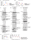
δ-Secretase-cleaved APP fragment selectively binds to CEBPB. (A) CEBPB-GFP was co-transfected with GST-tagged full-length APP, tau, α-syn or their δ-secretase-truncated fragments into HEK293 cells. In 48 h, GST pull-down was performed and analysed with anti-GFP antibody (first panel). The expression of pulled down GST-tagged fragments (second panel) and constructs in whole cell lysates (third and fourth panels) were examined. The pulled down proteins were quantified relative to the expression level of each construct in cell lysates. Scatter plot, mean ± SEM (*P <0.05, one-way ANOVA). (B) γ-Secretase and δ-secretase-derived CTFs show high binding activity with GST-CEBPB. GFP-tagged full-length APP or δ-secretase-, γ-secretase-, BACE1-derived CTF was co-transfected with GST-CEBPB into HEK293 cells. At 48 h, a GST pull-down assay was conducted and analysed with anti-GFP (first panel). Pulled-down GST-CEBPB is shown in the second panel and the expression of constructs in cell lysate are shown in the third and fourth panels. The bound APP fragments were quantified relative to the expression level of each construct. Scatter plot, mean ± SEM (*P <0.05, **P < 0.01, as compared with APP C586-695-GFP group, one-way ANOVA). (C) APP C586-695 selectively binds to CEBPB but neither to C/EBPα nor to HSF1 pointing to a highly specific interaction. The lower part shows the quantification of pulled down proteins that relative to the expression level of each construct. Scatterplot, mean ± SE; ***P <0.001, one-way ANOVA. (D) Endogenous APP C586-695 but not APP FL associates with endogenous CEBPB in 3xTg mice. Brain lysates from 12-month-old 3xTg and 3xTg/AEP KO mice were immunoprecipitated with IgG, anti-APP C586 or anti-APP C, respectively. The co-immunoprecipitation bound proteins were analysed with anti-CEBPB (first panel of left part). Pulled down APP C586 fragments are also shown in the second panel of left part. Quantification of the bounded CEBPB is shown in the bottom panel. Scatter plot, mean ± SEM; ***P <0.001, one-way ANOVA. The expression levels of CEBPB, APP C586-695, APP FL in mouse brain lysates (right) are also shown. For immunoprecipitation, three mice were used in each group. (E) APP C586-695 strongly interacts with CEBPB in Alzheimer’s disease patient brains. Healthy control and Alzheimer’s disease patient brain lysates were immunoprecipitated with IgG or anti-APP C586 and analysed with anti-CEBPB (top) and anti-Fe65 (middle). Bottom: Pulled-down APP C586-695. For immunoprecipitation, three samples were used for each group. Right: Quantification of the bound CEBPB. Scatter plot, mean ± SEM; *P <0.05, one-way ANOVA. H.C. = heavy chain.
Figure 2
Figure 2
APP C586-695 fragment interacts with CEBPB in the nucleus. (A) APP C586-695 strongly associates with CEBPB and Fe65, but Fe65 does not interact with CEBPB in 3xTg mice. Brain lysates from 12-month-old 3xTg, 3xTg/AEP KO and 3xTg/CEBPB+/− mice were immunoprecipitated with control IgG and anti-APP C586 (top) or control IgG and anti-Fe65 (bottom), analysed with anti-CEBPB, anti-Fe65 for APP C586 pulled-down groups (top) or anti-CEBPB, anti-APP C586 for Fe65 pulled-down groups (bottom). L.C. = light chain. (B) APP C586-695 activates CEBPB via enhancing its phosphorylation by activating phospho-ERK1/2 signalling. The nuclear and cytoplasmic fractions of 12-month-old 3xTg and 3xTg/AEP KO mice brain tissues were prepared, then analysed by western blot. Right: Quantification of AEP, APPC586, CEBPB and p-CEBPB. Scatter plot, mean ± SEM; *P <0.05, **P <0.05, ***P <0.001, one-way ANOVA). (C) GST pull-down assay shows Fe65 promotes the association between APP C586-695 and CEBPB dose-dependently in HEK293 cells. GFP-CEBPB and GST-APP C586-695 were transfected with different amounts of Fe65-Myc into HEK293 cells. At 48 h, GST pull-down assay was conducted and analysed with anti-GFP (first panel, left). Pulled-down GST-APP C586-695 (second panel, left) and the expression of constructs in cell lysates (third and fourth panels) are as shown. The bound CEBPB-GFP was quantified relative to the expression level of construct in cell lysate (top panel, right). Scatter plot, mean ± SEM; **P <0.01, one-way ANOVA. ELISA shows that Fe65 promoted the production of amyloid-β dose-dependently in APP C586-695 and CEBPB co-transfected HEK293 cells (middle panel, right). Fe65 enhances LGMN promoter luciferase activity dose-dependently in HEK293 cells transfected with Fe65, CEBPB and APP 586-695 followed by OGD treatment (bottom panel, right). Data represent mean ± SEM of three independent experiments; *P <0.05, **P <0.01, one-way ANOVA. (D) Co-transfection assay indicates that Fe65 promotes the nuclear distribution of APP C586-695. Triple co-expression of APP C586-695, CEBPB and Fe65 in HEK293 cells resulted in prominent APP C586-695 nuclear residency. Scale bar = 20 µm. Right: Quantification of the ration of nuclear/cytoplasmic APP C586-695. Data are a box-and-whisker plots showing median, interquartile interval, minimum, maximum and all data-points. *P <0.05, **P <0.01, one-way ANOVA.
Figure 3
Figure 3
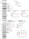
APP C586-695 escalates CEBPB transcriptional activity. (A) OGD significantly enhances LGMN promoter luciferase activity in cells transfected with CEBPB, which was further promoted in the presence of APP C586-695. AEP luciferase promoter, CEBPB and APP CTF plasmids were co-transfected into HEK293 cells. Upon OGD treatment and recovery, cells were collected and subjected to the luciferase assay. Data represent mean ± SEM of three independent experiments; *P <0.05, **P <0.01, one-way ANOVA. (B) Real-time PCR analysis reveals that both APP C586-695 and AICD significantly enhance CEBPB transcriptional activity at the LGMN mRNA level, and APP C586-695 but not AICD50 or APP FL increased MAPT mRNA transcription in SH-SY5Y cells with OGD treatment. Data represent mean ± SEM of three independent experiments; *P <0.05, one-way ANOVA. The expression of the constructs in A and B are validated in C. (D) Primary neuronal cultures were infected with GST-CEBPB and GFP, APP C586-695-GFP, C99-GFP or APP 1-695-GFP viruses at DIV5 and followed by the treatment of 2 µM pre-aggregated amyloid-β for 24 h at in vitro Day (DIV)13, then subjected to western blot. Immunoblotting assay shows that APP C586-695 exhibited stronger stimulatory effect than APP C99 or APP FL on CEBPB, leading to more abundant δ-secretase and δ-secretase-truncated tau and APP fragments. (E and F) Primary neuronal cultures were co-infected with shCon or shCEBPB and APP 586-695-GFP or APP FL-GFP viruses at DIV5, then followed by immunoblotting and qRT-PCR analysis at DIV13. Quantitative RT-PCR and immunoblotting assays revealed that APP C586-695 enhanced CEBPB transcriptional activity on LGMN, APP, MAPT and IL6 compared to APP FL, which are diminished by depletion of CEBPB. Data represent mean ± SEM of three independent experiments (*P <0.05, **P <0.01, one-way ANOVA).
Figure 4
Figure 4
Inhibition of APP cleavage by δ-secretase inhibits CEBPB activation by amyloid-β. (A) Primary cultured neurons were pre-treated with CP11 or dimethyl sulphoxide (DMSO) for 30 min then followed by the treatment of 2 µM pre-aggregated amyloid-β or DMSO for 24 h. Western blot (WB) analysis reveals that CP11 pretreatment diminished amyloid-β-induced δ-secretase activation and the subsequent cleavage of APP and tau. Right: Quantification of the active band of AEP. Data represent mean ± SEM; *P <0.05, one-way ANOVA). (B) AEP inhibitor CP11 inhibits the activity of δ-secretase. Data represent mean ± SEM of three independent experiments; *P <0.05, t-test). (C) Quantitative RT-PCR analysis shows that mRNA levels of Lgmn, App, and Mapt are reduced by CP11. Data represent mean ± SEM of three independent experiments (*P <0.05, **P <0.01, t-test). (D) ELISA shows that the inflammatory cytokines levels such as IL-6 and IL1-β are significantly decreased upon CP11 treatment. Data represent mean ± SEM of three independent experiments (*P <0.05, t-test). (E) Primary neuronal cultures were co-infected with shCon or shCEBPB and APPSLA or APPSLA N373AN585A viruses at DIV5, then followed by immunoblotting and qRT-PCR analysis at DIV13. APPSLA elicited CEBPB transcriptional activity on LGMN compared to δ-secretase-uncleavable APPSLA, which was diminished by depletion of CEBPB. Right: Quantification of the bands of CEBPB, AEP and APP (human). Data represent mean ± SEM (*P <0.05, one-way ANOVA). (F) Quantitative RT-PCR assay validates the knockdown efficiency of shCEBPB virus by detecting mRNA level of CEBPB. Data represent mean ± SEM of three independent experiments; *P <0.05, one-way ANOVA). (G) Quantitative RT-PCR assay reveals that mRNA levels of LGMN, APP, MAPT and IL6 are significantly decreased in neurons infected with δ-secretase-uncleavable APPSLA than wild-type APPSLA, and their mRNA level differences were diminished by depletion of CEBPB. Data represent mean ± SEM of three independent experiments (*P <0.05, **P <0.01, one-way ANOVA).
Figure 5
Figure 5
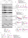
APP C586-695 but not APP FL stimulates CEBPB activities and promotes Alzheimer’s disease pathologies and cognitive disorders. (A) Hippocampal CA1 tissues from lentivirus-injected (GFP, APP C586-695-GFP, APP FL-GFP) wild-type (WT) or Cebpb+/− mice were analysed by western blot with various antibodies (n =4 mice/group). Immunoblotting analysis shows that the protein levels of δ-secretase and δ-secretase-truncated APP, tau fragments are higher in mice expressing APP C586-695 in brain tissue compared to control or APP FL in wild-type mice and this difference is attenuated in Cebpb+/− mice. Right: Quantification of the active band of AEP. Data are a box-and-whisker plots showing median, interquartile interval, minimum, maximum and all data-points; *P <0.05, one-way ANOVA. (B) Quantitative RT-PCR analysis reveals that mRNA levels of Lgmn, App, Mapt, Il6 are significantly enhanced by APP C586-695 compared with control or APP FL in wild-type mice and the mRNA levels are reduced in Cebpb+/− brains, but there is no significant difference between APP C586-695 and APP FL in Cebpb+/− mice. Data represent min to max and whiskers plots of four mice per group (*P <0.05, **P <0.01, one-way ANOVA). (C) ELISA shows that amyloid-β40 and amyloid-β42 concentrations are strongly enhanced by APP C586-695 in wild-type mice compared with APP FL and the difference is attenuated in Cebpb+/− mice. Data represent min to max and whiskers plots of five mice per group (*P <0.05, **P <0.01, one-way ANOVA). (D and E) Morris water maze (MWM) test (D) and conditional fear conditioning (CFC) analysis (E) of cognitive functions. APP C586-695 in CA1 elicited more learning and memory deficits than APP FL in wild-type mice and the effects were abolished in Cebpb+/− mice. Box-and-whisker plots, n =8 mice/group, *P <0.05, **P <0.01, one-way ANOVA).
Figure 6
Figure 6
Blockade of δ-secretase APP cleavage abrogates APP SLA mutant-induced amyloid pathologies and cognitive dysfunctions. (A) Hippocampal CA1 tissues from adeno-associated virus (AAV)-injected (Control, APPSLA, APPSLA N373A/N585A) wild-type or Cebpb+/− mice were analysed by western blot with various antibodies (n =4 mice/group). Immunoblotting analysis shows that the protein levels of δ-secretase and δ-secretase-truncated APP, tau fragments are higher in APPSLA-expressing brain samples than control or APPSLA N373A/N585A in wild-type mice and this difference is attenuated in Cebpb+/− mice. Right: Quantification of the active band of AEP. Data are box-and-whisker plots showing median, interquartile interval, minimum, maximum and all data-points (*P <0.05, one-way ANOVA). (B) Quantitative RT-PCR analysis reveals that mRNA levels of Lgmn, App, Mapt, Il6 are significantly enhanced by APPSLA compared with control or APPSLA N373A/N585A in wild-type mice. Levels of mRNA are reduced in Cebpb+/− brains but there is no significant difference between APPSLA and APPSLA N373A/N585A in Cebpb+/− mice. Data represent min to max and whiskers plots of four mice per group (*P <0.05, **P <0.01, one-way ANOVA). (C) ELISA shows that amyloid-β40 and amyloid-β42 concentrations are strongly enhanced by APPSLA in wild-type mice compared with APPSLA N373A/N585A and the difference is attenuated in Cebpb+/− mice. Data represent min to max and whiskers plots of five mice per group (*P <0.05, **P <0.01, one-way ANOVA). (D and E) Morris water maze (MWM) test (D) and conditional fear conditioning (CFC) analysis (E) of cognitive functions. The APPSLA in CA1 elicited more learning and memory deficits than APPSLA N373A/N585A in wild-type mice and the effects were abolished in Cebpb+/− mice. Box-and-whisker plots, n =8 mice/group, *P <0.05, one-way ANOVA).
Figure 7
Figure 7
Clearance of APP C586-695 from 5xFAD diminishes CEBPB transcriptional activity and rescues cognitive functions. (A) APP C586 antibody administration reduces the levels of APP C586 (first panel). APP C586 antibody attenuates CEBPB-stimulated δ-secretase activation and decreases the subsequent δ-secretase-truncated APP and tau fragment generation compared with IgG injection group. Four-month-old 5xFAD mice were treated with control IgG or anti-APP C586 via intraperitoneal injection (twice/week, 2 mg/kg) for 3 months. Hippocampus lysates were analysed by western blot (n =4 mice/group). Right: Quantification of the APP CTF, APP FL and amyloid-β. Box-and-whisker plots, *P <0.05, **P <0.01, ***P <0.001, t-test). (B) Quantitative RT-PCR reveals that mRNA levels of Lgmn and App are significantly diminished after anti-APP C586 treatment versus control IgG. Data represent min to max and whiskers plots of four mice per group (*P <0.05, **P <0.01, t-test). (C) ELISA shows that amyloid-β40 but not amyloid-β42 is significantly reduced upon APP C586 removal. Data represent min to max and whiskers plots of five mice per group (*P <0.05, t-test). (D and E) Morris water maze (D) and conditional fear conditioning (CFC) test (E) show that removal of APP C586 attenuates the learning and memory dysfunctions in 5xFAD mice (box-and-whisker plots, n =8 mice/group, *P <0.05, t-test). (F) Immunofluorescence and immunohistochemistry staining with anti-amyloid-β and Thioflavin-S staining reveal that APP C586 antibody administration reduces amyloid-β aggregates both in the cortex (scale bar = 300 µm) and the hippocampus (scale bar = 500 µm). Right: Quantification of the average number of plaques per slice and % area of plaques (plaque area/total area of analysed region). Plaque analysis data represent min to max and whiskers plots of six mice per group (*P <0.05, **P <0.01, t-test).

Similar articles

Cited by

References

    1. Selkoe DJ. Alzheimer's disease: A central role for amyloid. J Neuropathol Exp Neurol. 1994;53:438–447. - PubMed
    1. Estus S, Golde TE, Younkin SG.. Normal processing of the Alzheimer's disease amyloid beta protein precursor generates potentially amyloidogenic carboxyl-terminal derivatives. Ann NY Acad Sci. 1992;674:138–148. - PubMed
    1. Gersbacher MT, Goodger ZV, Trutzel A, Bundschuh D, Nitsch RM, Konietzko U.. Turnover of amyloid precursor protein family members determines their nuclear signaling capability. PLoS One. 2013;8:e69363. - PMC - PubMed
    1. McLoughlin DM, Miller CC.. The FE65 proteins and Alzheimer's disease. J Neurosci Res. 2008;86:744–754. - PubMed
    1. Cao X, Sudhof TC.. A transcriptionally [correction of transcriptively] active complex of APP with Fe65 and histone acetyltransferase Tip60. Science. 2001;293:115–120. - PubMed

Publication types

Substances