. 2021 Jan-Dec:30:9636897211009559.
doi: 10.1177/09636897211009559.

Multilayered Human Skeletal Muscle Myoblast Sheets Promote the Healing Process After Colonic Anastomosis in Rats

Affiliations

Multilayered Human Skeletal Muscle Myoblast Sheets Promote the Healing Process After Colonic Anastomosis in Rats

Takashi Nakamura et al. Cell Transplant. 2021 Jan-Dec.

Abstract

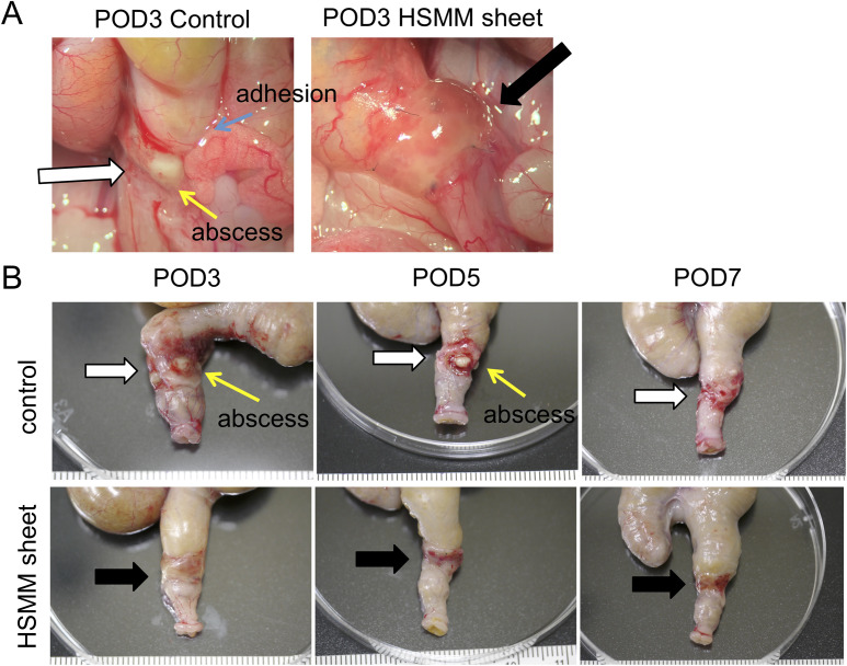
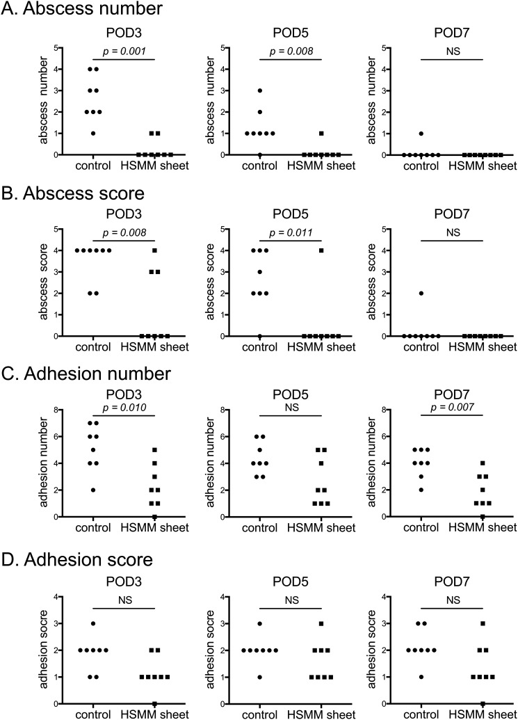
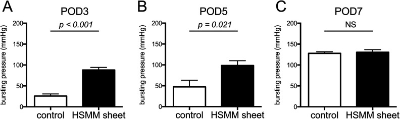
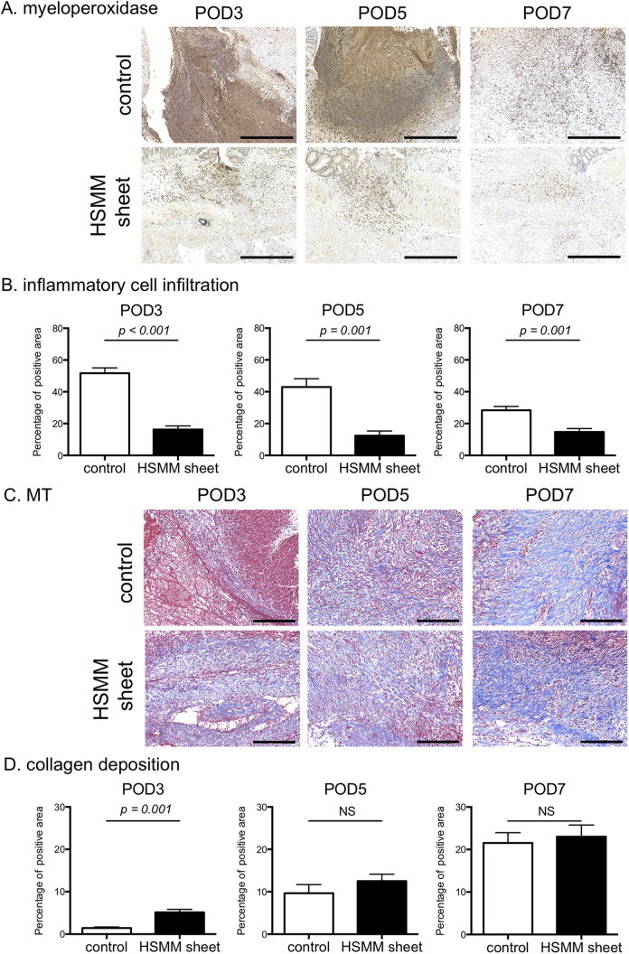
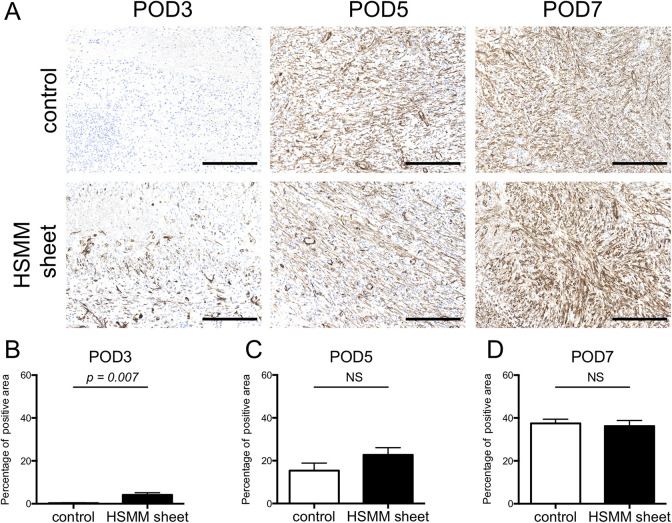
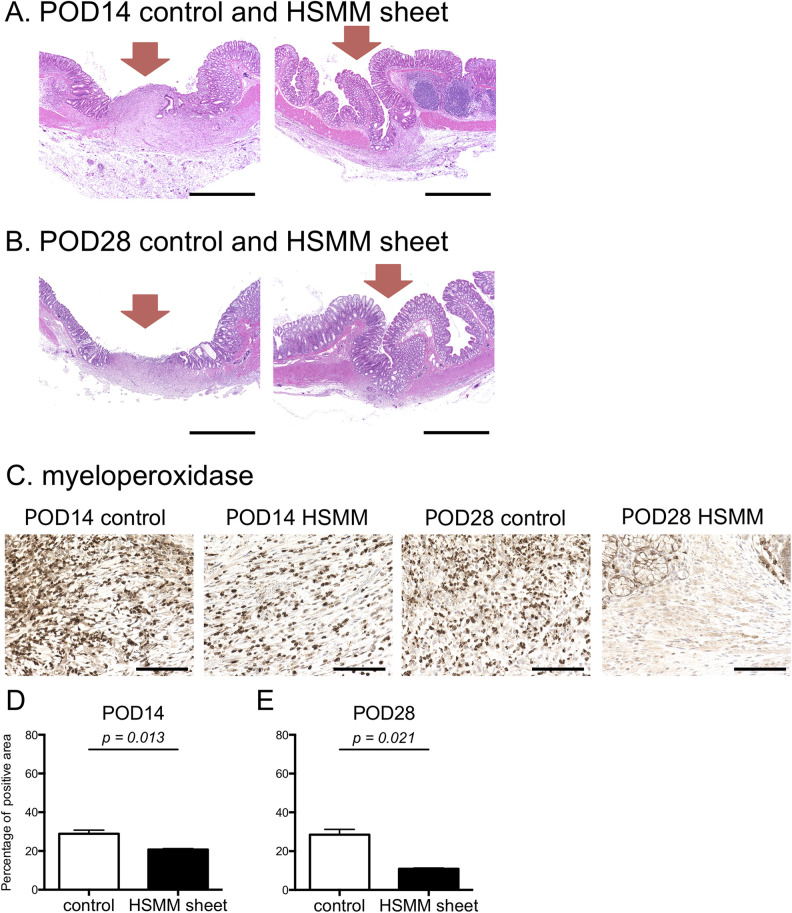
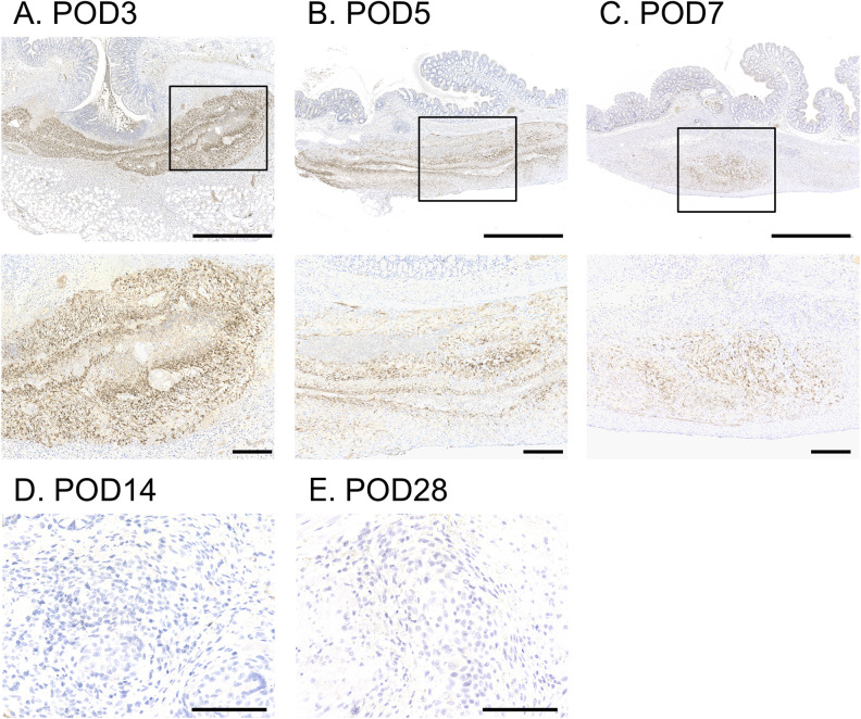
Colorectal anastomotic leakage is one of the most feared and fatal complications of colorectal surgery. To date, no external coating material that can prevent anastomotic leakage has been developed. As myoblasts possess anti-inflammatory capacity and improve wound healing, we developed a multilayered human skeletal muscle myoblast (HSMM) sheet by periodic exposure to supraphysiological hydrostatic pressure during repeated cell seeding. We assessed whether the application of an HSMM sheet can promote the healing process after colonic anastomosis. Partial colectomy and insufficient suturing were employed to create a high-risk colo-colonic anastomosis model in 60 nude rats. Rats were divided into a control group (n = 30) and an HSMM sheet group (n = 30). Macroscopic findings, anastomotic bursting pressure, and histology at the colonic anastomotic site were evaluated on postoperative day (POD) 3, 5, 7, 14, and 28. The application of an HSMM sheet significantly suppressed abscess formation at the anastomotic site compared to the control group on POD3 and 5. The anastomotic bursting pressure in the HSMM sheet group was higher than that in the control group on POD3 and 5. Inflammatory cell infiltration in the HSMM sheet group was significantly suppressed compared to that in the control group throughout the time course. Collagen deposition in the HSMM sheet group on POD3 was significantly abundant compared to that in the control group. Regeneration of the mucosa at the colonic anastomotic site was promoted in the HSMM sheet group compared to that in the control group on POD14 and 28. Immunohistochemical analysis demonstrated that surviving cells in the HSMM sheet gradually decreased with postoperative time and none were detected on POD14. These results suggest that the application of a multilayered HSMM sheet may prevent postoperative colonic anastomotic leakage.

Keywords: anastomotic leakage; hydrostatic pressure; mechanical stress; skeletal myoblast; tissue engineering.


Conflict of interest statement

Declaration of Conflicting Interests: The author(s) declared no potential conflicts of interest with respect to the research, authorship, and/or publication of this article.

Figures

Figure 1.
Figure 1.
Transplantation of HSMM sheets. (A) Hematoxylin & eosin (HE; left) and Masson’s trichrome stains (MT; right) of an HSMM sheet. Scale bars, 100 μm. (B-E) HSMM sheet transplantation procedure. After resection of the colonic segment (B), a full-thickness end-to-end anastomosis was created (C). The anastomotic site was wrapped with an HSMM sheet (D-E).
Figure 2.
Figure 2.
Representative images of macroscopic observation at the colonic anastomotic site. (A) Representative macroscopic images at the colonic anastomotic site after re-laparotomy on postoperative day (POD) 3 in the control group (left) and the HSMM sheet group (right). (B) Representative macroscopic images after resection of the anastomotic site on POD3 (left), 5 (middle), and 7 (right) in the control group (upper) and the HSMM sheet group (lower). White and black arrows indicate the anastomotic site and the transplanted HSMM sheet, respectively.
Figure 3.
Figure 3.
Macroscopic observations of the anastomotic site. (A) Abscess number, (B) abscess score, (C) adhesion number, and (D) adhesion score on postoperative day (POD) 3 (left), 5 (middle), and 7 (right). n = 8. Statistics were analyzed using Mann–Whitney U tests. NS, not significant.
Figure 4.
Figure 4.
Anastomotic bursting pressure on postoperative day (POD) 3 (A), 5 (B), and 7 (C). Results are presented as the mean ± SEM. n = 8. Statistics were analyzed using the unpaired t-test. NS, not significant.
Figure 5.
Figure 5.
Comparison of inflammatory cell infiltration and collagen deposition between the control group and the HSMM sheet group on postoperative day (POD) 3, 5, and 7. (A) Representative images of immunohistochemical stain for myeloperoxidase of the anastomotic site in the control (upper) and the HSMM sheet (lower) group on POD3 (left), 5 (middle), and 7 (right). Scale bars, 500 μm. (B) Inflammatory cell infiltration assessed by area percentage of myeloperoxidase-positive cells on POD3 (left), 5 (middle), and 7 (right). (C) Representative images of Masson’s trichrome (MT) stain of the anastomotic site in the control (upper) and the HSMM sheet (lower) group on POD3 (left), 5 (middle), and 7 (right). Scale bars, 200 μm. (D) Collagen deposition assessed by area percentage of collagen stained by MT stain on POD3 (left), 5 (middle), and 7 (right). Results are presented as the mean ± SEM. n = 8. Statistics were analyzed using the unpaired t-test. NS, not significant.
Figure 6.
Figure 6.
Expression level of α-SMA-positive cells at the anastomotic site on postoperative day (POD) 3, 5, and 7. (A) Representative images of immunohistochemical stain for α-SMA of the anastomotic site in the control (upper) and the HSMM sheet (lower) group on POD3 (left), 5 (middle), and 7 (right). Scale bars, 200 μm. (B-D) Comparison of area percentage of α-SMA-positive cells at the anastomotic site between the control and HSMM sheet group on POD 3 (B), 5 (C), and 7 (D). Results are presented as the mean ± SEM. n = 8. Statistics were analyzed using the unpaired t-test. NS, not significant.
Figure 7.
Figure 7.
Histological evaluation at the anastomotic site 14 and 28 days after transplantation of the HSMM sheet. (A, B) Representative images of hematoxylin & eosin stain in the control (left) and the HSMM sheet (right) group on postoperative day (POD) 14 (A) and 28 (B). There was discontinuation of the mucous membrane in the control group on POD14 and 28. Recovery of the continuity of the mucous membrane in the HSMM sheet group is evident on POD14 and 28. Scale bars, 1 mm. Red arrows indicate the anastomotic site. (C) Representative images of immunohistochemical stain for myeloperoxidase of the anastomotic site in the control and the HSMM sheet group on POD14 and 28. Scale bars, 100 μm. (D–E) Comparison of inflammatory cell infiltration at the anastomotic site on POD14 (D) and 28 (E). Results are presented as the mean ± SEM. n = 3. Statistics were analyzed using the unpaired t-test.
Figure 8.
Figure 8.
Remaining HSMM sheets at the anastomotic site. (A–C) Representative images of immunohistochemical stain for HLA on postoperative day (POD) 3 (A), 5 (B), and 7 (C). Upper images are low power field (scale bars, 1 mm). The selected area with the solid line is enlarged and illustrated below (scale bars, 200 μm). (D–E) Representative images of immunohistochemical stain for HLA of the anastomotic site on POD14 (D) and 28 (E). Scale bars, 100 μm. HLA-positive cells gradually diminished with POD, and were no longer detected on POD14 and 28.

Similar articles

Cited by

References

    1. Vogel JD, Eskicioglu C, Weiser MR, Feingold DL, Steele SR. The american society of colon and rectal surgeons clinical practice guidelines for the treatment of colon cancer. Dis Colon Rectum. 2017;60(10):999–1017. - PubMed
    1. Kingham TP, Pachter HL. Colonic anastomotic leak: risk factors, diagnosis, and treatment. J Am Coll Surg. 2009;208(2):269–278. - PubMed
    1. Snijders HS, Wouters MW, van Leersum NJ, Kolfschoten NE, Henneman D, de Vries AC, Tollenaar RA, Bonsing BA. Meta-analysis of the risk for anastomotic leakage, the postoperative mortality caused by leakage in relation to the overall postoperative mortality. Eur J Surg Oncol. 2012;38(11):1013–1019. - PubMed
    1. Walker KG, Bell SW, Rickard MJ, Mehanna D, Dent OF, Chapuis PH, Bokey EL. Anastomotic leakage is predictive of diminished survival after potentially curative resection for colorectal cancer. Ann Surg. 2004;240(2):255–259. - PMC - PubMed
    1. Law WL, Choi HK, Lee YM, Ho JW, Seto CL. Anastomotic leakage is associated with poor long-term outcome in patients after curative colorectal resection for malignancy. J Gastrointest Surg. 2007;11(1):8–15. - PubMed

Publication types

LinkOut - more resources