. 2021 May 28;84(5):1425-1433.
doi: 10.1021/acs.jnatprod.0c00840. Epub 2021 Apr 21.

Bufadienolides from the Eggs of the Toad Bufo bufo gargarizans and Their Antimelanoma Activities

Affiliations

Bufadienolides from the Eggs of the Toad Bufo bufo gargarizans and Their Antimelanoma Activities

Shi-Wen Zhou et al. J Nat Prod. .

Abstract

Toads produce potent toxins, named bufadienolides, to defend against their predators. Pharmacological research has revealed that bufadienolides are potential anticancer drugs. In this research, we reported nine bufadienolides from the eggs of the toad Bufo bufo gargarizans, including two new compounds (1 and 3). The chemical structures of 1 and 3, as well as of one previously reported semisynthesized compound (2), were elucidated on the basis of extensive spectroscopic data interpretation, chemical methods, and X-ray diffraction analysis. Compound 1 is an unusual 19-norbufadienolide with rearranged A/B rings. A biological test revealed that compounds 2 and 4-8 showed potent cytotoxic activities toward human melanoma cell line SK-MEL-1 with IC50 values less than 1.0 μM. A preliminary mechanism investigation revealed that the most potent compound, 8, could induce apoptosis via PARP cleavage, while 5 and 6 significantly suppressed angiogenesis in zebrafish. Furthermore, an in vivo biological study showed that 5, 6, and 8 inhibit SK-MEL-1 cell growth significantly.


Conflict of interest statement

The authors declare no competing financial interest.

Figures

Figure 1.
Figure 1.
Key HMBC and COSY correlations of 13.
Figure 2.
Figure 2.
Key NOE correlations of 1.
Figure 3.
Figure 3.
X-ray ORTEP drawing of 2.
Figure 4.
Figure 4.
Compounds 5, 6, and 8 induce the cleavage of PARP. SK-MEL-1 cells were treated with 5, 6, and 8 for 48 h. The protein expression level of PARP was examined by Western blotting. β-Actin was used as a loading control. Sorafenib (10 μM) was used as the positive control.
Figure 5.
Figure 5.
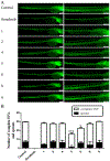
Compounds 5 and 6 exhibit significant antiangiogenic effects in vivo. Zebrafish embryos (Tg:Fli1-GFP) at 12 h postfertilization (hpf) were selected and treated with isolates (20 μM). At 24 hpf, the embryo membranous eggshells were broken. At 48 hpf, the development of embryonic blood vessels was observed under a fluorescence microscope (A), and the numbers of intersegmental vessels (ISVs) and sprout angiogenesis were counted (B). *P < 0.05, ***P < 0.01 versus control, one-way ANOVA, post hoc comparisons, Tukey, columns, mean; error bar, SD. Sorafenib (1 μM) was used as the positive control.
Figure 6.
Figure 6.
Compounds 5 and 6 have no obvious effect on transgenic zebrafish (Tg:Flk1-GFP). Zebrafish embryos at 12 h postfertilization (hpf) were selected and treated with 5 (20 μM) and 6 (20 μM). At 24 hpf, the embryo membranes were broken. At 48 hpf, the development of embryonic blood vessels was observed under a fluorescence microscope (A) and the numbers of intersegmental vessels (ISVs) and sprout angiogenesis were counted (B). One-way ANOVA, post hoc comparisons, Tukey, columns, mean; error bar, SD. Sorafenib (1 μM) was used as the positive control.
Figure 7.
Figure 7.
Compounds 5, 6, and 8 exert antimelanoma effects in vivo by CM-Dil staining. Melanoma cells SK-MEL-1 prestained by CM-Dil dye (2 μM) were microinjected into the yolk of zebrafish and treated with 5 (20 μM), 6 (20 μM), and 8 (10 μM) for 48 h. CM-Dil fluorescence was observed by microscopy. Sorafenib (0.5 μM) was used as the positive control. Left: CM-Dil-staining cells, middle: bright field, right: merge. The black oval area indicates the melanoma area.
Figure 8.
Figure 8.
Toxicity of 1, 5, 6, 8, and 9 in zebrafish larvae. Zebrafish larvae at 48 hpf were treated with compounds 1, 5, 6, 8, and 9 and then were observed by a stereomicroscope (Olympus SZX7) every 12 h. (A) Survival analysis on zebrafish larvae treated with compounds 1, 5, 6, 8, and 9. (B) Pathological changes in zebrafish larvae, including pericardial edema and curved tail, were observed under the light field of a stereomicroscope. The red arrow points to the pericardial edema and curved tail. (C) Percent of cardiac distortion in zebrafish larvae.

References

    1. Schmidt BR; Hodl W; Schaub M Ecology 2012, 93, 657–667. - PubMed
    1. Ujvari B; Casewell NR; Sunagar K; Arbuckle K; Wuster W; Lo N; O’Meally D; Beckmann C; King GF; Deplazes E; Madsen T Proc. Natl. Acad. Sci. U. S. A 2015, 112, 11911–11916. - PMC - PubMed
    1. Steyn PS; van Heerden FR Nat. Prod. Rep 1998, 15, 397–413. - PubMed
    1. Gao HM; Popescu R; Kopp B; Wang ZM Nat. Prod. Rep 2011, 28, 953–969. - PubMed
    1. Prassas I; Diamandis EP Nat. Rev. Drug Discovery 2008, 7, 926–935. - PubMed

Publication types

Substances