. 2021 Jun 3;81(11):2428-2444.e6.
doi: 10.1016/j.molcel.2021.03.044. Epub 2021 Apr 20.

FANCM regulates repair pathway choice at stalled replication forks

Affiliations

FANCM regulates repair pathway choice at stalled replication forks

Arvind Panday et al. Mol Cell. .

Abstract

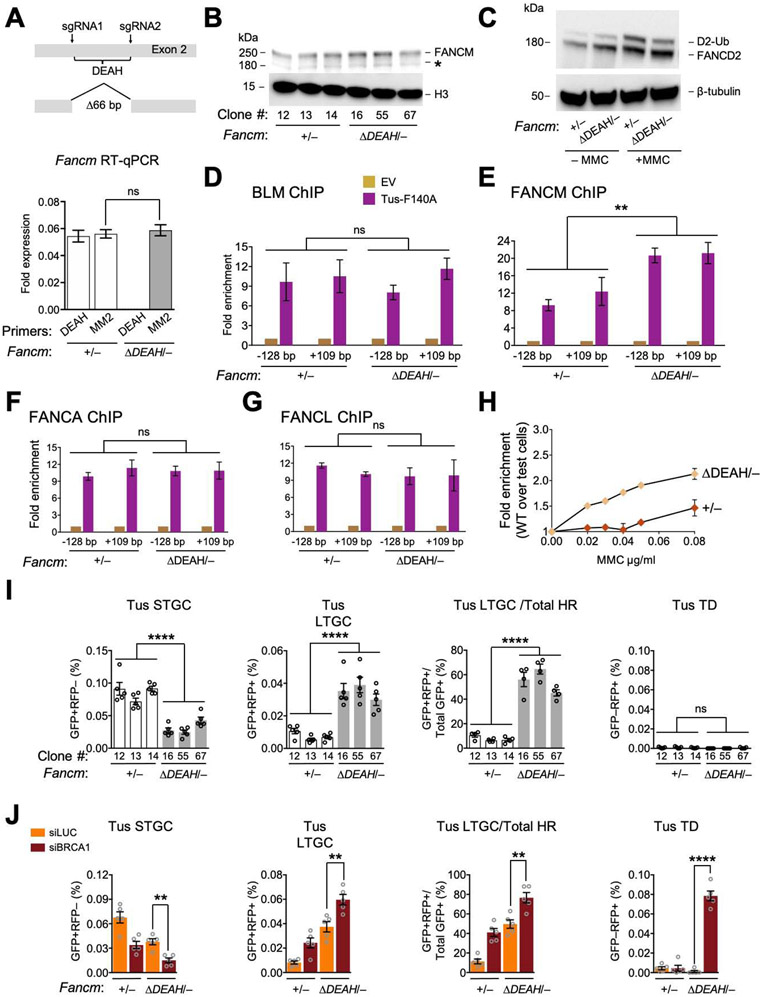
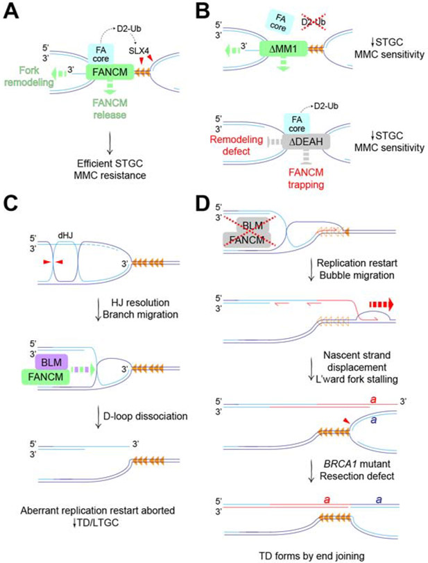
Repair pathway "choice" at stalled mammalian replication forks is an important determinant of genome stability; however, the underlying mechanisms are poorly understood. FANCM encodes a multi-domain scaffolding and motor protein that interacts with several distinct repair protein complexes at stalled forks. Here, we use defined mutations engineered within endogenous Fancm in mouse embryonic stem cells to study how Fancm regulates stalled fork repair. We find that distinct FANCM repair functions are enacted by molecularly separable scaffolding domains. These findings define FANCM as a key mediator of repair pathway choice at stalled replication forks and reveal its molecular mechanism. Notably, mutations that inactivate FANCM ATPase function disable all its repair functions and "trap" FANCM at stalled forks. We find that Brca1 hypomorphic mutants are synthetic lethal with Fancm null or Fancm ATPase-defective mutants. The ATPase function of FANCM may therefore represent a promising "druggable" target for therapy of BRCA1-linked cancer.

Keywords: BRCA1; Bloom’s syndrome helicase; FANCM; Fanconi anemia; break-induced replication; genomic instability; homologous recombination; replication restart; synthetic lethality; tandem duplication.


Conflict of interest statement

Declaration of interests The authors declare no competing interests.

Figures

Figure 1.
Figure 1.. FANCM is recruited to Tus/Ter-stalled mammalian replication forks.
See also Figure S1. A. Cartoon of FANCM protein and gene structure. DEAH: helicase domain. B. 6xTer-HR reporter and repair products of Tus/Ter-induced fork stalling. Grey boxes: mutant GFP alleles. Orange triangle: 6xTer array. Blue line: I-SceI restriction site. Ovals A and B: artificial 5’ and 3’ RFP exons. Red ovals: wild type RFP-coding exons. STGC/LTGC: short/long tract gene conversion. TD: tandem duplication. Red zig-zag: non-homologous TD breakpoint. C. FancmΔ85 allele, showing frame-shift product with premature stop codon (*). D. Left panel: Immunoblot of chromatin-extracted FANCM in Fancm+/+ and FancmΔ85/Δ clones. H3: Histone H3 loading control. Right panel: loss of FANCM band in siFANCM-treated samples. siLuc: control siRNA to Luciferase. *: background band. E. Immunoblot showing FANCD2 ubiquitination in Fancm+/+ and FancmΔ85/Δ clones in presence or absence of MMC. β-tubulin: loading control. F. Proliferative competition assay in MMC, measuring enrichment of GFP+ Fancm+/+ vs. GFP FancmΔ85/Δ cells. Data shows mean ± standard deviation (SD), n=3. Here and all subsequent growth assays, data normalized to 0 μg/mL MMC. G. ChIP analysis of FANCM at Tus/Ter. Cartoon shows qPCR primer positions for ChIP (red half-arrows; GFP sequence not shown). Numbers indicate distance in bp from outer primer to nearest edge of 6xTer array. Orange triangles: Ter sites. Blue line: I-SceI restriction site. Lower panel: FANCM ChIP 24 hours after transfection with empty vector (EV; gold) or Tus-F140A (purple). Data here and in all subsequent ChIP figures shows mean of 2−ΔΔCT values, normalized to EV and β-Actin control locus (see STAR methods). Data shows mean ± SD. Analysis by one-way ANOVA (n=3). In this and all subsequent figures: *: P < 0.05: **: P < 0.01; ***: P < 0.001; ****: P < 0.0001; ns: not significant. H. ChIP analysis of FANCM spreading at Tus/Ter or at I-SceI-induced DSB. Data shows mean ± SD (n=3).
Figure 2.
Figure 2.. FANCM regulates three distinct pathways of stalled fork repair.
See also Figure S2. A. Tus/Ter-induced repair in Fancm+/+ (white) clones vs. FancmΔ85/Δ (gray) clones. Data shows mean ± SEM. Analysis by one-way ANOVA (n=4). B. I-SceI-induced HR measured in same experiment. C. Representative raw FACS data (uncorrected for transfection efficiency) for Fancm+/+ and FancmΔ85/Δ cells co-transfected with empty vector (EV), I-SceI or Tus and siRNAs as shown. FACS plots pooled from n=4. Numbers show percentages. D. Tus/Ter-induced repair in Fancm+/+ clone #48 and FancmΔ85/Δ85 clone #39 co-transfected with Tus and siRNAs as shown (see STAR Methods). Data shows mean ± SEM. Analysis by Student’s t-test (n=4). E. I-SceI-induced repair measured in parallel in same experiment.
Figure 3.
Figure 3.. The FANCM-FA core complex interaction specifically mediates Tus/Ter-induced STGC.
See also Figure S3. A. FancmΔMM1 allele and RT qPCR analysis of MM1 and MM2 encoding mRNA in Fancm+/− and FancmΔMM1/− cells. Red half-arrows: RT qPCR primers. Here and in all subsequent expression analyses, data normalized to Gapdh mRNA using the 2−ΔCT method (see STAR methods). Data shows mean ± SD. Analysis by Student’s t-test (n=3). B. Immunoblot of chromatin-extracted FANCM in Fancm+/− and FancmΔMM1/− clones. *: non-specific band. C. Immunoblot showing FANCD2 ubiquitination in Fancm+/− and FancmΔMM1/− clones. D. Proliferative competition assay in MMC, measuring enrichment of GFP+ Fancm+/− vs. GFP FancmΔMM1/− cells. Data shows mean ± SD (n=3). E and F. ChIP analysis of FANCM (E) and FANCA, FANCL and BLM (F) at Tus/Ter in Fancm+/− and FancmΔMM1/− cells. Data shows mean ± SD. Analysis by one-way ANOVA (n=3). G. Tus/Ter-induced repair in Fancm+/− (white) clones vs. FancmΔMM1/− (gray) clones. Data shows mean ± SEM. Analysis by one-way ANOVA (n=5). H. Tus/Ter-induced repair in Fancm+/− vs. FancmΔMM1/− clones co-transfected with Tus and siRNAs as shown. Data shows mean ± SEM. Analysis by Student’s t-test (n=6).
Figure 4.
Figure 4.. The FANCM-BLM interaction suppresses LTGC and TD formation at stalled forks.
See also Figure S4. A. FancmΔMM2 allele and RT qPCR analysis of MM1 and MM2 encoding mRNA in Fancm+/− and FancmΔMM2/−clones. Data shows mean ± SD. Analysis by Student’s t-test (n=3). B. Immunoblot of chromatin-extracted FANCM in Fancm+/− and FancmΔMM2/− clones. C. Immunoblot showing FANCD2 ubiquitination in Fancm+/− and FancmΔMM2/− cells. D and E. ChIP analysis of BLM (D), FANCM, FANCA, and FANCL (E) at Tus/Ter in Fancm+/− and FancmΔMM2/− cells. Analysis by one-way ANOVA (n=3). F. Tus/Ter-induced HR in Fancm+/− (white) clones vs. FancmΔMM2/− (gray) clones. Data shows mean ± SEM. Analysis by one-way ANOVA (n=4). G. Tus/Ter-induced repair in Fancm+/− vs. FancmΔMM2/− clones co-transfected with Tus and siRNAs as shown. Data shows mean ± SEM. Analysis by Student’s t-test (n=5).
Figure 5.
Figure 5.. FANCM ATP hydrolysis mutants are defective for FANCM-mediated stalled fork repair.
See also Figures S5 and S6. A. FancmΔDEAH allele and RT qPCR analysis of MM2 and DEAH encoding mRNA in Fancm+/− and FancmΔDEAH/− clones. Data shows mean ± SD. Analysis by Student’s t-test (n=3). B. Immunoblot of chromatin-extracted FANCM in Fancm+/− and FancmΔDEAH/− clones. C. Immunoblot showing FANCD2 ubiquitination in Fancm+/− and FancmΔDEAH clones. D-G. ChIP analysis of BLM (D), FANCM (E), FANCA (F) and FANCL (G) at Tus/Ter in Fancm+/− and FancmΔDEAH/− cells. Data shows mean ± SD. Analysis by one-way ANOVA (n=3). H. Proliferative competition assay in presence of MMC, measuring enrichment of GFP+ Fancm+/− vs. GFP FancmΔDEAH/−cells (n=3). Error bars: SD. I. Tus/Ter-induced HR in Fancm+/− (white) clones vs. FancmΔDEAH/− (gray) clones. Data shows mean ± SEM. Analysis by one-way ANOVA (n=5). J. Tus/Ter-induced repair in Fancm+/− vs. FancmΔDEAH/− clones co-transfected with Tus and siRNAs as shown. Data shows mean ± SEM. Analysis by Student’s t-test (n=5).
Figure 6.
Figure 6.. Synthetic lethal interaction between Brca1 and Fancm mutations.
See also Figure S7. A. Left panel: wild type BRCA1 product of Brca1fl allele. Exon 11-encoded region shown. Cre converts Brca1fl to Brca1Δ, with in-frame deletion of exon 11. Right panel: Brca1 exons 10-12 in Brca1fl allele with PCR primers indicated (red half-arrows). Black triangles: loxP elements. B. Brca1fl/11 and Brca1Δ/11 PCR products using primers from panel A. C. RT qPCR analysis of Brca1 Exon 11-encoded mRNA in Brca1fl/11 and Brca1Δ/11 clones. Brca1 gene expression level was normalized to Gapdh using the 2−ΔCT method. Data shows mean ± SEM (n=3). D. Brca1fl/11 and Brca1Δ/11 allele recovery in unselected clones following Cre transduction of Brca1fl\11 cells carrying the Fancm genotypes shown.
Figure 7.
Figure 7.. Mechanisms of FANCM in stalled fork repair.
A. FANCM mediates error-free HR (i.e., STGC) at forks bidirectionally arrested at Tus/Ter (orange triangles). FANCM recruits FA core complex, promoting FANCD2 ubiquitination (D2-Ub) and SLX4-mediated incisions (red triangles). FANCM motor function (green dashed arrows) promotes fork remodeling and timely release of FANCM from stalled fork. B. Upper panel: FANCM ΔMM1 mutant retains motor functions but is defective for FA core complex recruitment and FANCD2 ubiquitination, resulting in reduced STGC and increased MMC sensitivity. Lower panel: FANCM ΔDEAH mutant retains FA core complex recruitment and FANCD2 ubiquitination. Defective fork remodeling, possibly combined with FANCM trapping, results in reduced STGC and increased MMC sensitivity. C. FANCM and BLM act in a concerted fashion to suppress aberrant replication fork restart. Hypothetical mechanism of D-loop formation at stalled fork in the absence of a strand exchange step. BLM can dissolve post-replicative double Holliday junction (dHJ). Alternative processing by HJ resolution (red triangles) generates D-loop at stalled fork, with accompanying sister chromatid exchange. Dashed light blue lines: resected nascent lagging strands at stalled fork. FANCM/BLM-mediated branch migration (green and purple dashed arrow) dissociates D-loop, preventing aberrant fork restart. D. Defects in FANCM/BLM interaction allow D-loop to persist, favoring resumption of nascent leading strand synthesis (red half-arrow) with displacement of Tus from Ter (empty orange triangles). Engagement of unknown helicase(s) (red dashed arrow) extends aberrant replication restart by bubble migration. This mechanism is BIR-like but, as shown in C, might not be break-induced. Leftward normal fork duplicates genomic segment ‘a’ bounded, at one end, by the site of fork stalling and, at the other end, by the site of displacement of the restarted leading strand (red). BRCA1 loss impairs DNA end resection, preventing collapse of TD back to single copy by single strand annealing. By default, tandem duplication forms by end joining.

References

    1. Adamo A, Collis SJ, Adelman CA, Silva N, Horejsi Z, Ward JD, Martinez-Perez E, Boulton SJ, and La Volpe A (2010). Preventing nonhomologous end joining suppresses DNA repair defects of Fanconi anemia. Mol Cell 39, 25–35. - PubMed
    1. Amunugama R, Willcox S, Wu RA, Abdullah UB, El-Sagheer AH, Brown T, McHugh PJ, Griffith JD, and Walter JC (2018). Replication Fork Reversal during DNA Interstrand Crosslink Repair Requires CMG Unloading. Cell reports 23, 3419–3428. - PMC - PubMed
    1. Bachrati CZ, Borts RH, and Hickson ID (2006). Mobile D-loops are a preferred substrate for the Bloom's syndrome helicase. Nucleic Acids Res 34, 2269–2279. - PMC - PubMed
    1. Bakker ST, van de Vrugt HJ, Rooimans MA, Oostra AB, Steltenpool J, Delzenne-Goette E, van der Wal A, van der Valk M, Joenje H, te Riele H, et al. (2009). Fancmdeficient mice reveal unique features of Fanconi anemia complementation group M. Human molecular genetics 18, 3484–3495. - PubMed
    1. Bogliolo M, Bluteau D, Lespinasse J, Pujol R, Vasquez N, d'Enghien CD, Stoppa-Lyonnet D, Leblanc T, Soulier J, and Surrallés J (2018). Biallelic truncating FANCM mutations cause early-onset cancer but not Fanconi anemia. Genetics in medicine : official journal of the American College of Medical Genetics 20, 458–463. - PubMed

Publication types

MeSH terms

LinkOut - more resources