. 2021 Apr 21;13(8):11678-11695.
doi: 10.18632/aging.202861. Epub 2021 Apr 21.

Piccolo is essential for the maintenance of mouse retina but not cochlear hair cell function

Affiliations

Piccolo is essential for the maintenance of mouse retina but not cochlear hair cell function

Peipei Li et al. Aging (Albany NY). .

Abstract

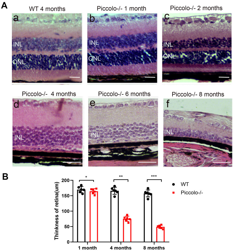
Piccolo is a presynaptic protein with high conservation among different species, and the expression of Piccolo is extensive in vertebrates. Recently, a small fragment of Piccolo (Piccolino), arising due to the incomplete splicing of intron 5/6, was found to be present in the synapses of retinas and cochleae. However, the comprehensive function of Piccolo in the retina and cochlea remains unclear. In this study, we generated Piccolo knockout mice using CRISPR-Cas9 technology to explore the function of Piccolo. Unexpectedly, whereas no abnormalities were found in the cochlear hair cells of the mutant mice, significant differences were found in the retinas, in which two layers (the outer nuclear layer and the outer plexiform layer) were absent. Additionally, the amplitudes of electroretinograms were significantly reduced and pigmentation was observed in the fundoscopy of the mutant mouse retinas. The expression levels of Bassoon, a homolog of Piccolo, as well as synapse-associated proteins CtBP1, CtBP2, Kif3A, and Rim1 were down-regulated. The numbers of ribbon synapses in the retinas of the mutant mice were also reduced. Altogether, the phenotype of Piccolo-/- mice resembled the symptoms of retinitis pigmentosa (RP) in humans, suggesting Piccolo might be a candidate gene of RP and indicates Piccolo knockout mice are a good model for elucidating the molecular mechanisms of RP.

Keywords: CRISPR/Cas9; cochlea; mouse; piccolo; retina.


Conflict of interest statement

CONFLICTS OF INTEREST: The authors declare that they have no conflicts of interest.

Figures

Figure 1
Figure 1
Generation of Piccolo-/- mouse. (A) Schematic diagram of the CRISPR/Cas9 knockout strategy. (B) Schematic illustration of Cas9-mediated targeting of the mouse Piccolo gene (sgRNA and PAM sequences are underlined) and comparison of Piccolo DNA sequences in wild type and Piccolo-/- (4 bp deletion) mice. (C) Schematic illustration of the frameshift mutation in Piccolo-/- mice. The altered amino acid sequence is marked in red. “*” indicates a premature stop codon. The translation of protein is terminated at the point of the arrow. Also shown are the Piccolo wild type protein sequence and domain organization. (D) Sequencing chromatograms of Piccolo in wild type (WT) mice, heterozygous (HET; Piccolo+/-) mice, and homozygous (Homo; Piccolo-/-) mice. The red box indicates the bases missing in the homozygous (Homo) Piccolo-/- mice. (E) Relative Piccolo expression in Piccolo-/- and WT mice as measured by Q-PCR. (F) Western blot analysis of Piccolo protein in the retina of Piccolo-/- and WT mice. GADPH was used as the loading control.
Figure 2
Figure 2
Auditory function of Piccolo-/- mice. (A) Gross morphology of Piccolo-/- and wild type (WT) mice at the age of one month (P30). Scale bar = 2cm. (B) Overall appearance of cochleae from Piccolo-/- and WT mice at P30. Scale bar = 15mm. (C) H&E staining of cochlear sections in Piccolo-/- and WT mice at P30. Scale bar = 20 μm. (D) Scanning electron microscopic images of cochlear hair bundles in Piccolo-/- and WT mice at P30. Scale bar = 5 μm. (EG) ABR thresholds of Piccolo-/- (n = 3) and WT (n = 3) mice (at the age of one month, 4 months and 8 months, respectively) to click stimuli and to 4, 8, 16, and 32 kHz stimuli. Error bars = Means ± SD. (H) Representative ABR recording from control (n = 5) and Piccolo-/- (n = 3) mice at P30. The asterisk indicates a significant difference between WT and Piccolo-/- mice. *P < 0.05, **P < 0.01, by two-tailed Student’s t test. Error bars = Mean ± SD.
Figure 3
Figure 3
Structure and function of retina in wild type and Piccolo-/- mouse. (A) Eyes from wild type (WT) and Piccolo-/- mice. (B) Mean diameters of eyeballs from WT (n = 8) and Piccolo-/- (n = 8) mice. (C) ERG recording a-wave amplitudes from WT (n=6) and Piccolo-/- (n=6) mice at P30. (D) ERG recording b-wave amplitudes from WT (n=6) and Piccolo-/- (n=6) mice at P30. (E) ERG recording a-wave amplitudes from WT (n=6) and Piccolo-/- (n=6) mice at 4 months. (F) ERG recording b-wave amplitudes from WT (n=6) and Piccolo-/- (n=6) mice at 4 months. The asterisk indicates a significant difference between WT and Piccolo-/- mice. *P < 0.05, **P < 0.01, by two-tailed Student’s t test. Error bars = Mean ± SD.
Figure 4
Figure 4
Retinal anatomy in Piccolo-/- mice. (A) H&E stained paraffin sections of retinas from Piccolo-/- and wild type (WT) mice. The image below represents the magnification of the upper image. (B) Immunofluorescence staining with DAPI, α-PKC (marker for bipolar cells), and anti-Piccolo antibodies in the retina of Piccolo-/- and WT mice. Scale bars: 20 μm.
Figure 5
Figure 5
Retinal anatomy in wild type and Piccolo-/- mice. (A) H&E stained sections of retinas from Piccolo-/- and wild type mice at one, two, four, six and eight months of age, respectively. (B) The thickness of retina in Piccolo-/- and wild type (WT) mice (n = 6 per group) at 1, 4 and 8 months; ***P < 0.001, by two-tailed Student’s t test.
Figure 6
Figure 6
Examination of the retinal fundus in Piccolo-/- mice. (A) Optical coherence tomography (OCT) of the retina in Piccolo-/- mice compared with the wild type (WT) mice at P30; Scale bars: 100 μm. (B) Retinal fundus imaging in one-month-old WT and Piccolo-/- mice.
Figure 7
Figure 7
The morphology, location, and numbers of ribbon synapses in Piccolo-/- mice. (A) Immunostaining with anti-CtBP2 antibodies of retina cryosections from wild type (WT) and Piccolo-/- mice at P30. DAPI stains nuclei. Scale bars: 20 μm. (B) The morphology of ribbon synapses in the Piccolo-/- and WT mice. Scale bars: 10 μm. (C) The cytoplasmic location of retinal ribbon synapses (indicated by arrows) in Piccolo-/- mice. Scale bars: 1 μm.
Figure 8
Figure 8
Expression levels of synaptic ribbon-related proteins in Piccolo-/- mice. (A) Relative mRNA levels of ribbon-related genes in Piccolo-/- and wild type (WT) mice. n=5. (B) Western blot analysis of CtBP2 in Piccolo-/- and WT mice (GADPH was used as the loading control.) n=5. (C) Relative CtBP2 band intensities normalized to GADPH. n=5. (D) Relative mRNA levels of Bassoon in Piccolo-/- and WT mice. n=5. (E) Western blot analysis of Bassoon in Piccolo-/- and WT mice. (GADPH was used as the loading control.) n=5. (F) Relative Bassoon band intensities normalized to GADPH. Data represent the means ± SD. **P < 0.01, ***P < 0.001, by two-tailed Student’s t test, n=5.

References

    1. Moser T, Grabner CP, Schmitz F. Sensory processing at ribbon synapses in the retina and the cochlea. Physiol Rev. 2020; 100:103–44. 10.1152/physrev.00026.2018 - DOI - PubMed
    1. Zhai RG, Vardinon-Friedman H, Cases-Langhoff C, Becker B, Gundelfinger ED, Ziv NE, Garner CC. Assembling the presynaptic active zone: A characterization of an active one precursor vesicle. Neuron. 2001; 29:131–43. 10.1016/s0896-6273(01)00185-4 - DOI - PubMed
    1. Gundelfinger ED, Fejtova A. Molecular organization and plasticity of the cytomatrix at the active zone. Curr Opin Neurobiol. 2012; 22:423–30. 10.1016/j.conb.2011.10.005 - DOI - PubMed
    1. Zanazzi G, Matthews G. The molecular architecture of ribbon presynaptic terminals. Mol Neurobiol. 2009; 39:130–48. 10.1007/s12035-009-8058-z - DOI - PMC - PubMed
    1. Südhof TC. The presynaptic active zone. Neuron. 2012; 75:11–25. 10.1016/j.neuron.2012.06.012 - DOI - PMC - PubMed

Publication types

MeSH terms