Case Reports
. 2021 Apr 21;14(4):e237662.
doi: 10.1136/bcr-2020-237662.

Chronic myeloid leukaemia-associated retinopathy

Affiliations
Case Reports

Chronic myeloid leukaemia-associated retinopathy

Ashish Khalsa et al. BMJ Case Rep. .

Abstract

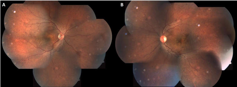
A 38-year-old man presented with mild blurring of vision in both eyes for the past 1 week. On examination, the retinal vessels were dilated and tortuous, along with multiple dot blot haemorrhages all over the fundus with yellowish white focal retinal infiltrates at the macula temporal to the fovea. The salmon pink discolouration of the blood column made us look at the peripheral blood smear, which was suggestive of chronic myeloid leukaemia, leading to a diagnosis of leukaemic retinopathy in both the eyes.

Keywords: macula; retina.


Conflict of interest statement

Competing interests: None declared.

Figures

Figure 1
Figure 1
Coloured fundus images of the right (A) and left (B) eyes showing dilated and tortuous vessels with a salmon pink hue (black arrow), multiple superficial retinal haemorrhages (white arrow) and white colour retinal infiltrates (blue arrow).
Figure 2
Figure 2
Fundus fluorescein angiogram of both eyes showing blocked fluorescence corresponding with the retinal haemorrhages and infiltrates in the arteriovenous phase (yellow arrows in A and B). There are pin point hyperfluorescence secondary to leak from the infiltrates as well as elsewhere in the late venous phase (red arrows in C and D). There is segmentation of the dye suggestive of stasis of blood flow in either eyes more prominent in the early frames (green arrow in B).
Figure 3
Figure 3
Optical coherence tomogram with transfoveal line scans in right (A) and left (B) eyes showing neurosensory detachment.
Figure 4
Figure 4
Optical coherence tomogram with line scan passing across the retinal infiltrate showing an increase in reflectivity of the inner retinal layers and loss of retinal architecture.
Figure 5
Figure 5
Coloured fundus photo montage of right (A) and left (B) eyes on follow-up showing resolution of retinal infiltrates, haemorrhages and return of normal colour of the retinal vessels.

References

    1. Zhou H, Xu R. Leukemia stem cells: the root of chronic myeloid leukemia. Protein Cell 2015;6:403–12. 10.1007/s13238-015-0143-7 - DOI - PMC - PubMed
    1. Soman S, Kasturi N, Srinivasan R, et al. . Ocular manifestations in leukemias and their correlation with hematologic parameters at a tertiary care setting in South India. Ophthalmol Retina 2018;2:17–23. 10.1016/j.oret.2017.05.009 - DOI - PubMed
    1. Rane PR, Barot RK, Gohel DJ, et al. . Chronic myeloid leukaemia presenting as bilateral retinal haemorrhages with multiple retinal infiltrates. J Clin Diagn Res 2016;10:ND04–5. 10.7860/JCDR/2016/18215.7822 - DOI - PMC - PubMed
    1. Rosenthal AR. Ocular manifestations of leukemia. A review. Ophthalmology 1983;90:899–905. 10.1016/S0161-6420(83)80013-X - DOI - PubMed

Publication types

LinkOut - more resources