. 2021 Apr 21;7(17):eabe0143.
doi: 10.1126/sciadv.abe0143. Print 2021 Apr.

Deformable microparticles for shuttling nanoparticles to the vascular wall

Affiliations

Deformable microparticles for shuttling nanoparticles to the vascular wall

Margaret B Fish et al. Sci Adv. .

Abstract

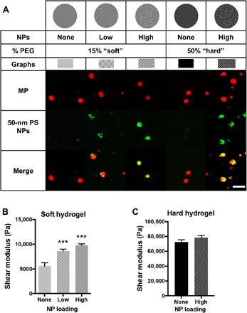
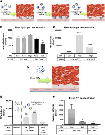
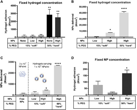
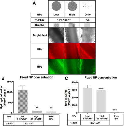
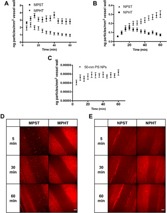
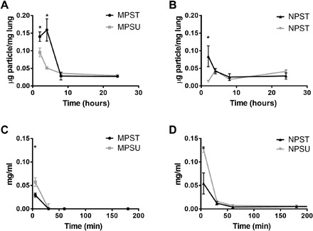
Vascular-targeted drug carriers must localize to the wall (i.e., marginate) and adhere to a diseased endothelium to achieve clinical utility. The particle size has been reported as a critical physical property prescribing particle margination in vitro and in vivo blood flows. Different transport process steps yield conflicting requirements-microparticles are optimal for margination, but nanoparticles are better for intracellular or tissue delivery. Here, we evaluate deformable hydrogel microparticles as carriers for transporting nanoparticles to a diseased vascular wall. Depending on microparticle modulus, nanoparticle-loaded poly(ethylene glycol)-based hydrogel microparticles delivered significantly more 50-nm nanoparticles to the vessel wall than freely injected nanoparticles alone, resulting in >3000% delivery increase. This work demonstrates the benefit of optimizing microparticles' efficient margination to enhance nanocarriers' transport to the vascular wall.


Figures

Fig. 1
Fig. 1. Material properties of NP-loaded hydrogel MPs.
(A) Schematic and representative confocal microscopy fluorescence images of hydrogel MPs evaluated, having varied modulus and NP loading. Red is MP hydrogel, green is 50-nm PS NPs, and the two are overlaid to show colocalization of NPs and hydrogel MPs. Scale bar, 5 μm. Swollen shear moduli for (B) 15% PEG and (C) 50% PEG hydrogels showing the influence of adding NPs to bulk material rheometry. Statistical analyses were performed using one-way analysis of variance (ANOVA) with Fisher’s least significant difference (LSD) test, where (***) indicates P < 0.001 in comparison to the nonloaded hydrogels. N = 3. Error bars plot SE.
Fig. 2
Fig. 2. Adhesion of NP-loaded hydrogel MPs to an inflamed HUVEC monolayer at 200 s−1 WSR.
(A) Schematic detailing “fixed MP concentration” in vitro flow experiments. Quantified (B) adhesion for anti–ICAM-1–coated hydrogel MPs dosed in blood at a fixed MP concentration and scaled to (C) the corresponding number of NPs delivered by the adherent hydrogel MPs in (B). (D) Schematic of the free NP in vitro flow experiments. (E) Number of NPs delivered to the vascular wall by free anti–ICAM-1–coated PS NPs dosed at 3 × 10⁷ NPs/ml or based on (F) the adhesion of hydrogel MPs dosed in blood to carry a fixed three times lower NP cargo of 1 × 10⁷ NPs/ml. For all, adhesion was quantified after 5 min of laminar blood flow over an IL-1β–activated HUVEC monolayer. N ≥ 3 human blood donors per particle condition. Statistical analysis of adherent density was performed using one-way ANOVA with Fisher’s LSD test, where (*) indicates P < 0.05, (**) indicates P < 0.01, (***) indicates P < 0.001, and (****) indicates P < 0.0001 versus the first bar in each plot. Error bars plot SE.
Fig. 3
Fig. 3. Adhesion of NP-loaded hydrogel MPs to an inflamed HUVEC monolayer at 1000 s−1 WSR.
Quantified (A) adhesion for hydrogel MPs dosed in blood at a fixed MP concentration and scaled to (B) the corresponding number of NPs delivered by the adherent hydrogel MPs in (A). (C) Number of NPs delivered to the vascular wall by free anti–ICAM-1–coated NPs dosed at 3 × 10⁷ NPs/ml or based on (D) the adhesion of hydrogel MPs dosed in blood to carry a fixed three times lower NP cargo of 1 × 10⁷ NPs/ml. For all, adhesion was quantified after 5 min of laminar blood flow over an IL-1β–activated HUVEC monolayer. N ≥ 3 human blood donors per particle condition. Statistical analysis of adherent density was performed using one-way ANOVA with Fisher’s LSD test, where (*) indicates P < 0.05, (**) indicates P < 0.01, (***) indicates P < 0.001, and (****) indicates P < 0.0001 versus the first bar in each plot. Error bars represent SE.
Fig. 4
Fig. 4. Delivery of NPs to an inflamed mesentery endothelium as a function of loading into hydrogel MPs.
(A) Representative bright-field and fluorescence images of particle adhesion to inflamed mesentery. n/a, not applicable. (B) Quantified adhesion density of three different particle conditions, 15% PEG, low loading hydrogel MPs, 15% PEG, high loading hydrogel MPs, and free NPs. Particles were dosed by equivalent NP payload. (C) Data scaled to the number of NPs delivered by adherent hydrogel MPs to show the efficiency of NP delivery by each VTC system. N = 3 mice per group, and statistical analysis was performed using one-way ANOVA with Fisher’s LSD test, where (**) indicates P < 0.01 and (***) indicates P < 0.001 compared to the low NP–loaded 15% PEG. Error bars plot SE. Scale bar, 50 μm.
Fig. 5
Fig. 5. Intravital microscopy analysis of MP and NP adhesion over time.
The adhesion of particles on an inflamed mesentery vein for (A) 2-μm PEG, (B) 500-nm PEG, and (C) 50-nm PS particles over an hour. Error bar represents SE for N = 3. Representative images of mesentery adhesion of (D) 2-μm and (E) 500-nm PEG NPs at 5, 30, and 60 min after particle injection. MPST, targeted soft MP; MPHT, targeted hard MP; NPST, targeted soft NP; NPHT, targeted hard NP. Scale bar, 50 μm.
Fig. 6
Fig. 6. Behavior of targeted hydrogel particles in mice with acute lung injury.
Accumulation of PEG-based (A) 2-μm MPs and (B) 500-nm NPs in lung injury mouse lungs 2, 4, 8, and 24 hours after particle injection. (C and D) Blood circulation profile over time in lung injury mice showing the concentration of PEG-based particles remaining in the bloodstream of lung injury mice minutes after particle injection. Plots are shown for both ICAM-1 targeted (T) and untargeted (U) particles. Bars represent the SE for N = 4. Statistical analysis was performed using one-way ANOVA with Fisher’s LSD test, where (*) indicates P < 0.05 compared to the untargeted particle at that time point.

References

    1. Thompson A. J., Mastria E. M., Eniola-Adefeso O., The margination propensity of ellipsoidal micro/nanoparticles to the endothelium in human blood flow. Biomaterials 34, 5863–5871 (2013). - PubMed
    1. Thompson A. J., Eniola-Adefeso O., Dense nanoparticles exhibit enhanced vascular wall targeting over neutrally buoyant nanoparticles in human blood flow. Acta Biomater. 21, 99–108 (2015). - PubMed
    1. Safari H., Kelley W. J., Saito E., Kaczorowski N., Carethers L., Shea L. D., Eniola-Adefeso O., Neutrophils preferentially phagocytose elongated particles—An opportunity for selective targeting in acute inflammatory diseases. Sci. Adv. 6, eaba1474 (2020). - PMC - PubMed
    1. Fish M. B., Fromen C. A., Lopez-Cazares G., Golinski A. W., Scott T. F., Adili R., Holinstat M., Eniola-Adefeso O., Exploring deformable particles in vascular-targeted drug delivery: Softer is only sometimes better. Biomaterials 124, 169–179 (2017). - PMC - PubMed
    1. Nel A. E., Madler L., Velegol D., Xia T., Hoek E. M., Somasundaran P., Klaessig F., Castranova V., Thompson M., Understanding biophysicochemical interactions at the nano-bio interface. Nat. Mater. 8, 543–557 (2009). - PubMed

Publication types