. 2021 Jun 10;95(13):e0217720.
doi: 10.1128/JVI.02177-20. Epub 2021 Jun 10.

Species-Specific Valid Ternary Interactions of HIV-1 Env-gp120, CD4, and CCR5 as Revealed by an Adaptive Single-Amino Acid Substitution at the V3 Loop Tip

Affiliations

Species-Specific Valid Ternary Interactions of HIV-1 Env-gp120, CD4, and CCR5 as Revealed by an Adaptive Single-Amino Acid Substitution at the V3 Loop Tip

Takaaki Koma et al. J Virol. .

Abstract

Molecular interactions of the variable envelope gp120 subunit of HIV-1 with two cellular receptors are the first step of viral infection, thereby playing pivotal roles in determining viral infectivity and cell tropism. However, the underlying regulatory mechanisms for interactions under gp120 spontaneous variations largely remain unknown. Here, we show an allosteric mechanism in which a single gp120 mutation remotely controls the ternary interactions between gp120 and its receptors for the switch of viral cell tropism. Virological analyses showed that a G310R substitution at the tip of the gp120 V3 loop selectively abolished the viral replication ability in human cells, despite evoking enhancement of viral replication in macaque cells. Molecular dynamics (MD) simulations predicted that the G310R substitution at a site away from the CD4 interaction site selectively impeded the binding ability of gp120 to human CD4. Consistently, virions with the G310R substitution exhibited a reduced binding ability to human lymphocyte cells. Furthermore, the G310R substitution influenced the gp120-CCR5 interaction in a CCR5-type dependent manner as assessed by MD simulations and an infectivity assay using exogenously expressed CCR5s. Interestingly, an I198M mutation in human CCR5 restored the infectivity of the G310R virus in human cells. Finally, MD simulation predicted amino acid interplays that physically connect the V3 loop and gp120 elements for the CD4 and CCR5 interactions. Collectively, these results suggest that the V3 loop tip is a cis-allosteric regulator that remotely controls intra- and intermolecular interactions of HIV-1 gp120 for balancing ternary interactions with CD4 and CCR5. IMPORTANCE Understanding the molecular bases for viral entry into cells will lead to the elucidation of one of the major viral survival strategies, and thus to the development of new effective antiviral measures. As shown recently, HIV-1 is highly mutable and adaptable in growth-restrictive cells, such as those of macaque origin. HIV-1 initiates its infection by sequential interactions of Env-gp120 with two cell surface receptors, CD4 and CCR5. A recent epoch-making structural study has disclosed that CD4-induced conformation of gp120 is stabilized upon binding of CCR5 to the CD4-gp120 complex, whereas the biological significance of this remains totally unknown. Here, from a series of mutations found in our extensive studies, we identified a single-amino acid adaptive mutation at the V3 loop tip of Env-gp120 critical for its interaction with both CD4 and CCR5 in a host cell species-specific way. This remarkable finding could certainly provoke and accelerate studies to precisely clarify the HIV-1 entry mechanism.

Keywords: CCR5; CD4; Env-gp120; HIV-1; V3 loop; adaptive mutation; in silico structural analysis; species specificity.


Figures

FIG 1
FIG 1
Genome organization and Env-gp120 glycoprotein. (A) Genome structures of HIV-1 clones used in this study and the domain structure of gp120. Genome organization of authentic HIV-1 (NL4-3) (49), prototype CCR5-tropic HIV-1mt (NL-DT562) (26), and newly constructed HIV-1 (NL-562-EX) clones are presented. Black and blue boxes indicate regions derived from SIVmac (SIVmac239) (61), and HIV-1 (SF162) (62), respectively. The enlarged gp120 region is presented. Constant (C1 to C5) and variable (V1 to V5) domains in gp120 are shown by white and orange areas, respectively. Four adaptive mutations in the V1/V2 loop of gp120 that we previously identified (27) are indicated. (B) Amino acid sequences of the V3 loops. Amino acid sequences of the V3 loops from parental 562 and its mutants carrying a single-amino-acid adaptive mutation are aligned. Numbers indicate amino acid positions in the Env proteins of HXB2 (GenBank M38432) and 562 (26) as shown. Bold letters in the 562 line show the residues for which we identified an adaptive mutation (27). The V3 tip and stem regions are indicated by blue and black lines/letters, respectively. Orange letters show the GPGR motif. The alignment was performed by Genetyx ver.14.
FIG 2
FIG 2
Structure of HIV-1 Env-gp120. (A) Overall architecture of a full-length HIV-1 Env gp120 trimer model in the unliganded state (27). The V1/V2 and V3 loops are highlighted in red and blue, respectively. Arrows indicate the locations of the V3 loop tips in the gp120 trimer. The locations of the V3 tip residues that were analyzed in the present study are shown by yellow and blue spheres. (B) Overall (left) and enlarged (right) views of a full-length HIV-1 Env gp120 monomer model in the CD4 and CCR5 bound state. The model was constructed by homology modeling using the cryo-EM structure of the HIV-1 CD4-gp120-CCR5 ternary complex (PDB code 6MEO) (20), followed by MD simulation for 100 ns (see the Materials and Methods for details). In the enlarged view, brown and poppy red spheres indicate amino acid residues at the V3 tip (G308 and G310) and those in CCR5 (K171 and I198), respectively. G308 and G310 residues are present in the conserved GPG triplet residues at the V3 tip. K171 and I198 residues are located at the ECL2 and TM5 domains in CCR5, respectively. These residues differ between human and CyM CCR5s.
FIG 3
FIG 3
Replication properties of parental 562 and its Env variant clones carrying an adaptive mutation. (A and B) Growth kinetics in lymphocyte cells. The schematic of Env-gp120 organization from Fig. 1 is presented at the top for clarity. Viruses prepared from 293T cells transfected with the indicated proviral clones were inoculated into CyM HSC-F (A) and human MT4/R5 (B) cells, and virus replication was monitored by the virion-associated RT activity in the culture supernatants. Equal amounts of viruses (2 × 106 RT units) were used for inoculation into HSC-F cells (2 × 105 cells). For the spin-infection of MT4/R5 cells (1 × 105 cells), equal amounts of viruses (6 × 106 RT units) were used. Representative data from three independent experiments are shown. Sample symbols in panel (B) are the same as those in panel (A). (C) Infectivity of 562 and G310R in TZM-bl cells. Viruses were inoculated into TZM-bl cells as described in the Materials and Methods, and 2 days postinfection, cells were lysed for luciferase assays. Viral infectivity relative to that for 562 in cells inoculated with 20 RT units (kcpm) was calculated by relative light units in cell lysates. Mean values ± standard errors (SEs) are shown (n = 3).
FIG 4
FIG 4
Effect of mutations in the V3 tip region on virus replication. (A) Frequency of amino acids at position 310 of Env-gp120 (by 562 numbering). Authentic (blue), adaptive (green), and selected (orange) amino acids are shown. Env sequences in the HIV-1 subtype B population (n = 19,419) were obtained from the HIV Sequence Database (http://www.hiv.lanl.gov/content/sequence/HIV/mainpage.html) (27). (B) Infection experiments of 562, 562EX, and their V3 mutants. The env sequences of 562 and 562EX are identical, and Env mutant clones (S304G, G308V/E, and G310R/L/E/K) in the context of 562 and 562 EX were constructed. Viruses prepared from 293T cells transfected with the indicated proviral clones were inoculated into HSC-F (upper) and MT4/R5 (lower) cells, and virus replication was monitored by the virion-associated RT activity in the culture supernatants. HSC-F cells (2 × 105 cells) and MT4/R5 cells (1 × 105 cells) were infected with equal amounts of viruses (5 × 105 RT units for HSC-F and 2 × 105 RT units for MT4/R5). Infection of MT4/R5 cells was performed by the spin-infection method. Representative data from three independent experiments are shown.
FIG 5
FIG 5
MD simulations of CD4-562 Env-gp120-CCR5 ternary complexes embedded in a lipid bilayer. (A) The CD4-gp120-CCR5 complex models were constructed by homology modeling and MD simulations at 1 bar and at 310 K for 100 ns in 0.15 M NaCl. Complex structures at 100 ns of MD simulations are shown. Individual complexes are viewed from the same direction by aligning along human CCR5s in the lipid bilayer. (B and C) The RMSDs of the receptor binding surfaces during 0 to 100 ns of MD simulations were calculated using the cpptraj module in AmberTools 16 as described previously (27, 36). (B) RMSDs of the gp120 regions for CD4 binding (37) during the MD simulations. The binding regions are composed of amino acid residues T246 to T251, Q359 to M369 (CD4 binding loop), T445 to E421, and R460 to D465 in 562 gp120. (C) RMSDs of the V3 loop (C294 to C328) of the 562 gp120 regions for CCR5 binding (20). (D and E) The RMSFs of the receptor binding surfaces during 90 to 100 ns of MD simulations were calculated using the ptraj module (60) in AmberTools 16 (27, 34–36). (D) RMSFs of the gp120 regions for CD4 binding (37). The regions indicated as a, b, c, and d are T246 to T251, Q359 to M369, T445 to E421, and R460 to D465 in 562 gp120 (26, 27), respectively. (E) RMSFs of the V3 loop (C294 to C328) of the 562 gp120 regions for CCR5 binding (20).
FIG 6
FIG 6
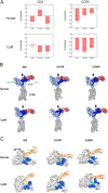
Effects of single-amino acid substitutions at the 562 Env-gp120 V3 tip on interactions of gp120 with CD4 and CCR5. (A) Binding free energies. The CD4-gp120-CCR5 ternary complex models from independent all-atom MD trajectories of the last 5 ns after 100 ns of MD simulations were used to calculate the binding free energies of the indicated receptor molecules using MMPBSA.py (59) in AmberTools16 (AMBER 2016, University of California, San Francisco). Mean values ± SEs are shown (n = 100). (B) Overall views of the ternary complex structures. The structures during 50 to 100 ns of MD simulations (n = 10) were superposed with CCR5, and the side views are shown. Arrows indicate the CD4 apex region for gp120 binding. (C) Top views of the ternary complexes at 100 ns of MD simulations. Location of the gp120 bridging sheets (37) and the V1/V2 region are indicated by cyan and blue, respectively.
FIG 7
FIG 7
Effect of the G310R mutation on the viral ability to bind to CyM HSC-F and human MT4/R5 cells. (A) CD4 expression levels on the HSC-F and MT4/R5 cell surfaces. Cells were stained with FITC-labeled CD4 antibody and analyzed by a flow cytometer. Numbers in the graphs indicate the mean fluorescence intensity of cell surface CD4. Negative controls are shown by black lines. (B) Viral ability to bind to HSC-F and MT4/R5 cells. Virus binding assays were performed as described in the Materials and Methods. Relative virus binding ability (Gag-p24 for EX-G310R/Gag-p24 for 562EX) is presented. Mean values ± SEs from three independent experiments are shown. Significance relative to control 562EX was determined by the Welch’s t test; ns, not significant.
FIG 8
FIG 8
Effect of various CCR5 expression levels on the infectivity of 562EX and 562EX-G310R (EX-G310R). (A) Alignment of human and CyM CCR5 amino acid sequences. Amino acid sequences from TM4 to TM5 in CCR5 (based on GPCRdb ccr5_human, https://gpcrdb.org/) are shown. TM4, ECL2, and TM5 regions are as indicated. Red letters show the different residues between CyM and human CCR5s. Alignment was performed by Genetyx ver.14. (B) The CCR5 expression level on the cell surface. MAGI cells were transfected with the indicated CCR5 expression vectors and, at 24 h posttransfection, cells were stained with anti-human CCR5 antibody for FACS analysis. Mean values ± SEs (n = 6) are shown. Significance was evaluated by the likelihood-ratio chi-squared test (n = 6); ns, not significant. (C) Infectivity of 562EX and 562EX-G310R in various CCR5-expressing cells. Viruses were prepared from 293T cells transfected with the proviral clones, and equal amounts of viruses were inoculated into MAGI cells transiently expressing various CCR5s. On day 2 postinfection, cells were lysed for beta-galactosidase assays. Viral infectivity relative to that for 562EX in CyM CCR5-expressing cells is shown. Mean values ± SEs (n = 5) are shown. Significance was determined by the Welch’s t test (n = 5); ns, not significant. Hu, human; KR, K171R; IM, I198M; KR/IM, K171R/I198M.
FIG 9
FIG 9
Molecular interactions among CD4, 562 Env-gp120, and CCR5. (A) Enlarged views of the human CD4-gp120 interaction site in the ternary complexes at 100 ns of MD simulation. Spheres indicate residues involved in hydrogen bonding in the 562 gp120 complex. The V3 loop, bridging sheets, and CD4 are indicated by light green, cyan, and light pink, respectively. (B) Enlarged views of the binding surface between human CCR5 and the HIV-1 gp120 V3 loop. A ternary complex structure composed of 562 gp120, human CD4, and human CCR5 at 100 ns of MD simulation (Fig. 5A, upper left panel) was used to address molecular interactions. The model suggests that the V3 loop is stably embedded in the CCR5 N-terminal pocket via hydrophobic interactions between the ITI residues immediately upstream of the V3 loop tip (amino acid numbers 305 to 307 in 562 gp120) and two hydrophobic cores of CCR5 (green dotted circles). The interactions involve connections between V3 I305 and CCR5 F182/W190 and between V3 I307 and CCR5 C178/C101. Note that CCR5 I198 and V3 G310 are located near CCR5 Q194 around the CCR5 hydrophobic core and V3 T306 in the ITI motif, respectively. Views from two arbitrary angles are shown.
FIG 10
FIG 10
Effect of G310R mutation on the growth ability of various virus clones. Amino acid sequences in the V3 loop regions of CCR5-tropic HIV-1mt clone 562 and the other two HIV-1mt clones designated B3AD8 and CI3 (63) are shown at the top. Dots indicate amino acids identical to those of 562. Note the boxed ITI motif. The growth abilities in macaque cells of the parental and mutant clones are qualitatively shown as follows: ++, parental clone; +++, grew better than each parental clone; +, grew more poorly than each parental clone; −, no detectable growth. For details, see Fig. 4 of this study and references and .

Similar articles

Cited by

References

    1. Chen B. 2019. Molecular mechanism of HIV-1 entry. Trends Microbiol 27:878–891. 10.1016/j.tim.2019.06.002. - DOI - PMC - PubMed
    1. Clapham PR, McKnight A. 2002. Cell surface receptors, virus entry and tropism of primate lentiviruses. J Gen Virol 83:1809–1829. 10.1099/0022-1317-83-8-1809. - DOI - PubMed
    1. Freed EO, Martin MA. 2013. Human immunodeficiency viruses: replication, p 1502–1560. In Knipe DM, Howley PM (ed), Fields virology, 6th ed, vol 2. Lippincott Williams & Wilkins, Philadelphia, PA.
    1. Wilen CB, Tilton JC, Doms RW. 2012. HIV: cell binding and entry. Cold Spring Harb Perspect Med 2:a006866. 10.1101/cshperspect.a006866. - DOI - PMC - PubMed
    1. Brandenberg OF, Magnus C, Rusert P, Regoes RR, Trkola A. 2015. Different infectivity of HIV-1 strains is linked to number of envelope trimers required for entry. PLoS Pathog 11:e1004595. 10.1371/journal.ppat.1004595. - DOI - PMC - PubMed

Publication types

LinkOut - more resources