. 2022 Jan;77(1):47-57.
doi: 10.1136/thoraxjnl-2020-216469. Epub 2021 Apr 21.

Sphingosine kinase 1 regulates lysyl oxidase through STAT3 in hyperoxia-mediated neonatal lung injury

Affiliations

Sphingosine kinase 1 regulates lysyl oxidase through STAT3 in hyperoxia-mediated neonatal lung injury

Alison W Ha et al. Thorax. 2022 Jan.

Abstract

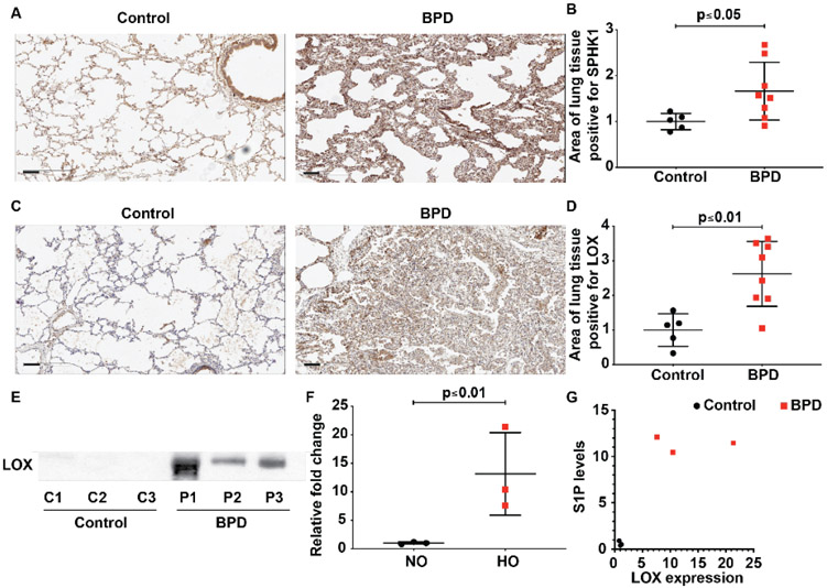
Introduction: Neonatal lung injury as a consequence of hyperoxia (HO) therapy and ventilator care contribute to the development of bronchopulmonary dysplasia (BPD). Increased expression and activity of lysyl oxidase (LOX), a key enzyme that cross-links collagen, was associated with increased sphingosine kinase 1 (SPHK1) in human BPD. We, therefore, examined closely the link between LOX and SPHK1 in BPD.

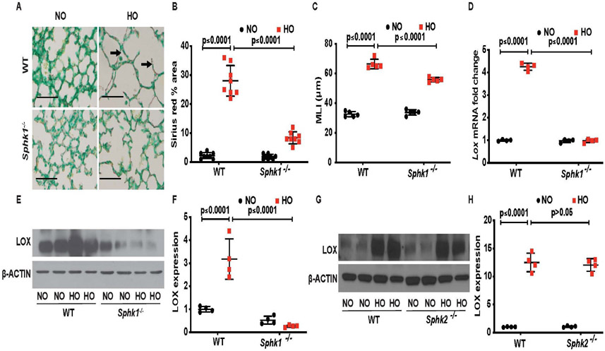
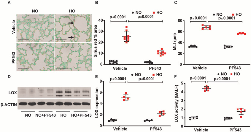
Method: The enzyme expression of SPHK1 and LOX were assessed in lung tissues of human BPD using immunohistochemistry and quantified (Halo). In vivo studies were based on Sphk1-/- and matched wild type (WT) neonatal mice exposed to HO while treated with PF543, an inhibitor of SPHK1. In vitro mechanistic studies used human lung microvascular endothelial cells (HLMVECs).

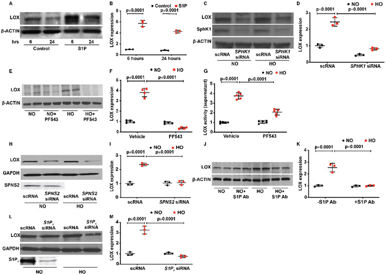
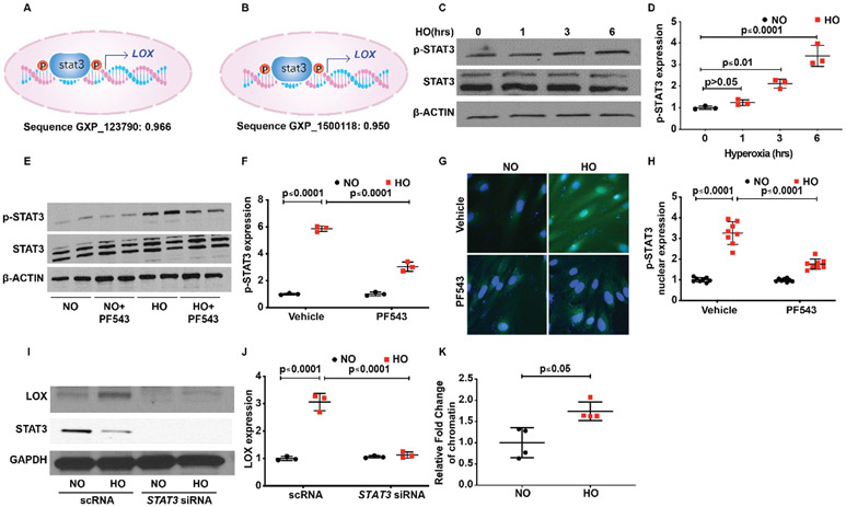
Results: Both SPHK1 and LOX expressions were increased in lungs of patients with BPD. Tracheal aspirates from patients with BPD had increased LOX, correlating with sphingosine-1-phosphate (S1P) levels. HO-induced increase of LOX in lungs were attenuated in both Sphk1-/- and PF543-treated WT mice, accompanied by reduced collagen staining (sirius red). PF543 reduced LOX activity in both bronchoalveolar lavage fluid and supernatant of HLMVECs following HO. In silico analysis revealed STAT3 as a potential transcriptional regulator of LOX. In HLMVECs, following HO, ChIP assay confirmed increased STAT3 binding to LOX promoter. SPHK1 inhibition reduced phosphorylation of STAT3. Antibody to S1P and siRNA against SPNS2, S1P receptor 1 (S1P1) and STAT3 reduced LOX expression.

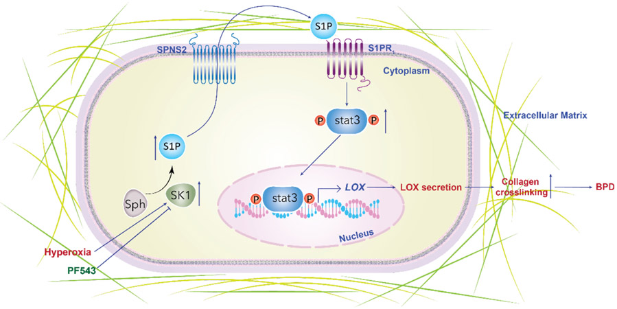
Conclusion: HO-induced SPHK1/S1P signalling axis plays a critical role in transcriptional regulation of LOX expression via SPNS2, S1P1 and STAT3 in lung endothelium.

Keywords: long term oxygen therapy (LTOT); oxidative stress; paediatric interstitial lung disease; paediatric lung disaese.


Conflict of interest statement

Competing interests: None declared.

Figures

Figure 1
Figure 1
Elevated sphingosine kinase 1 (SPHK1) and lysyl oxidase (LOX) expression are noted in lung tissue of neonates with severe BPD. Paraffin-embedded lung sections were used to assess expression of SPHK1 (A) and LOX by immunohistochemistry (C) in the lung tissue of neonates with BPD compared with controls. Intensity of antibody staining (brown) was quantified in halo system using scanned lung images (B and D). LOX was also elevated in the tracheal aspirates of newborns with severe BPD (E). Qquantified data show significance (F). Correlation test showed significant relationship (r=0.831) between LOX content in TA detected by immunoblot and the S1P level estimated (G). Original view, 10×, scale bar 100 μm. Statistical analyses done with Pearson correlation and Mann-Whitney U test. Details of patients are given in online supplemental tables 12. BPD, bronchopulmonary dysplasia; S1P, sphingosine-1-phosphate.
Figure 2
Figure 2
Deletion of sphingosine kinase 1 gene (Sphk1−/−) protects alveolarisation of murine neonatal lungs under hyperoxia (HO) accompanied by reduced collagen deposition/cross-linking. Representative H&E photomicrographs of lung sections from neonatal wild type (WT) and Sphk1−/− mice, exposed to room air or hyperoxia (HO) stained with sirius red for collagen (dark arrow). Sphk1−/− KO exposed to HO showed improved alveolarisation accompanied by reduced sirius red staining (A). The objective assessment of sirius red staining of the lungs was determined by quantification using ImageJ system. Following exposure to HO, sirius red staining of the lung of Sphk1−/− newborn mice is significantly lower compared with WT control exposed to HO (B) accompanied by protection against HO-induced lung injury as evidenced by improved mean linear intercept suggestive of improved alveolarisation (C). RT-PCR showed an increase in expression of Lox following HO in the WT, which was not seen in the Sphk1−/− exposed to HO (D). Whole lung tissue lysates subjected to SDS-PAGE and western blotting showed increased expression of LOX following HO in WT compared with no controls. This increase in expression was significantly reduced in the Sphk1−/− KO exposed to HO (E and F). Western blotting showed increased expression of LOX following HO in Sphk2−/− compared with no controls quite similar to the expression seen in WT (G and H). Original view, 40×, scale bar 100 μm. Statistical analyses done with two-way analysis of variance test, N 6–8/group, equal number of males and females used.
Figure 3
Figure 3
Inhibition of sphingosine kinase 1 protects alveolarisation of murine neonatal lungs under hyperoxia (HO) accompanied by reduced collagen deposition/cross-linking. Representative H&E photomicrographs of sirius red stained (dark arrow) lung sections from neonatal wild type (WT) mice, exposed to RA or HO and treated with PF543 or vehicle. WT pups exposed to HO but treated with PF543 showed improved alveolarisation accompanied by reduced sirius red staining (A). Quantified data of sirius red staining of the lungs were determined using ImageJ system. Following exposure to HO, sirius red staining of the lung of PF543-treated newborn mice is significantly lower compared with WT control exposed to HO (B). This was accompanied by protection against HO induced lung injury as evidenced by improved MLI suggestive of improved alveolarisation (C). Whole lung tissue lysates subjected to SDS-PAGE and western blotting showed increased expression of lysyl oxidase (LOX) following HO in WT compared with no controls. This increase in expression was significantly reduced in the WT exposed to HO and treated with PF543 (D and E). The reduction of expression of LOX in lung tissue lysates in the western blot of HO exposed and PF543 treated was accompanied by reduced LOX activity in the bronchoalveolar lavage fluid (F). Original view, 40×, scale bar 100 μm. Statistical analyses done with two-way analysis of variance test, N 6–8/group, equal number of males and females used.
Figure 4
Figure 4
Interruption of the SPHK1/S1P signalling pathway attenuates HO-induced increased LOX expression in human primary lung microvascular endothelial cells (HLMVECs). HLMVECs were used to study the influence of SPHK1/S1P signalling on LOX production. Exogenous S1P added to HLMVECs resulted in an increased expression of LOX at 6 hours, and there was no further increase noted at 24 hours (A and B). HO-induced increased expression of LOX at 6 hours was reduced by SPHK1 siRNA (C and D). Similar to the effect of siRNA, SPHK1 inhibitor, PF543, also prevented a HO-induced increase in LOX expression. No difference was noted between the two groups pretreated with or without PF543 (E and F). In consonance with a reduction of HO-induced expression of LOX following PF543 therapy, there was a significant reduction of LOX activity in the cell culture supernatant as well following PF543 therapy and exposure to HO (G). S1P transporter SPNS2 transporting S1P from inside the cell to the outside enabling it to bind to S1P receptors. siSPNS2 reduced HO-induced increased expression of LOX (H and I). S1P binding antibodies were added to the medium prior to exposure of HLMVECs to HO. Following exposure to HO, S1P antibody added group showed a reduced expression of LOX suggesting a role for S1P transported out of the cell into the medium in regulating LOX expression (J and K). siS1P1 reduced HO-induced increased expression of LOX compared with HO and NO controls (L and M). Statistical analyses done with two-way analysis of variance test. Images for control and treatment groups in microscopy experiments were collected at the same time under the same conditions. HO, hyperoxia; LOX, lysyl oxidase; NO, normoxia; S1P, sphingosine-1-phosphate receptor 1; SPHK1, sphingosine kinase 1.
Figure 5
Figure 5
Transcription factor STAT3 serves to activate S1P-mediated LOX transcription leading to HO-induced increased LOX expression in human primary lung microvascular endothelial cells (HLMVECs). In silico analyses showed a strong probability for STAT3 binding to the promoter site of LOX with a score of 0.966 and 0.95 (A and B). HLMVECs exposed to HO showed activation of STAT3 at 3 hours of HO as evidenced by its translocation to the nucleus, which was inhibited by PF543 the SPHK1 inhibitor (C and D; G &and H). HO induced an increase in expression of pSTAT3, which peaked at 6 hours. PF543 inhibited HO-induced increased expression of pSTAT3 at 6 hours (E and F). siSTAT3 inhibited HO-induced increased expression of LOX (I and J). Chromatin immune precipitation assay was done after 3 hours of HO, which showed a 1.7-fold increase in enrichment of STAT3 in the LOX promoter region. HO, hyperoxia; LOX, lysyl oxidase; NO, normoxia; S1P, sphingosine-1-phosphate receptor 1.
Figure 6
Figure 6
Schema showing the summary of the theoretical model of SPHK1/S1P/STAT3 axis in regulating LOX HO-induced experimental BPD. HO causes an increase in SPHK1, which stimulates the formation of S1P. HO leads to increased production of LOX leading to increased collagen cross-linking as evidenced by increased sirius red staining driven by SPHK1, S1P, SPNS2, S1P receptor 1 and STAT3 pathway. Inhibition of this pathway along its various components was associated with reduced LOX expression. Inhibition of SPHK1 was associated with reduced LOX production and collagen staining of lung tissue accompanied by restoration of lung alveolarisation. BPD, bronchopulmonary dysplasia; HO, hyperoxia; LOX, lysyl oxidase; S1P, sphingosine 1 phosphate; SPHK1, sphingosine kinase 1.

Similar articles

Cited by

References

    1. Andresen JH, Saugstad OD. Oxygen metabolism and oxygenation of the newborn. Semin Fetal Neonatal Med 2020;25:101078. - PubMed
    1. Bhandari V Hyperoxia-derived lung damage in preterm infants. Semin Fetal Neonatal Med 2010;15:223–9. - PMC - PubMed
    1. Kallet RH, Matthay MA. Hyperoxic acute lung injury. Respir Care 2013;58:123–41. - PMC - PubMed
    1. Ware LB, Matthay MA. The acute respiratory distress syndrome. N Engl J Med 2000;342:1334–49. - PubMed
    1. Xu X, Han M, Li T, et al. Effective treatment of severe COVID-19 patients with tocilizumab. Proc Natl Acad Sci U S A 2020;117:10970–5. - PMC - PubMed

Publication types

Substances