. 2021 Apr 27;118(17):e2016363118.
doi: 10.1073/pnas.2016363118.

HIM-17 regulates the position of recombination events and GSP-1/2 localization to establish short arm identity on bivalents in meiosis

Affiliations

HIM-17 regulates the position of recombination events and GSP-1/2 localization to establish short arm identity on bivalents in meiosis

Saravanapriah Nadarajan et al. Proc Natl Acad Sci U S A. .

Abstract

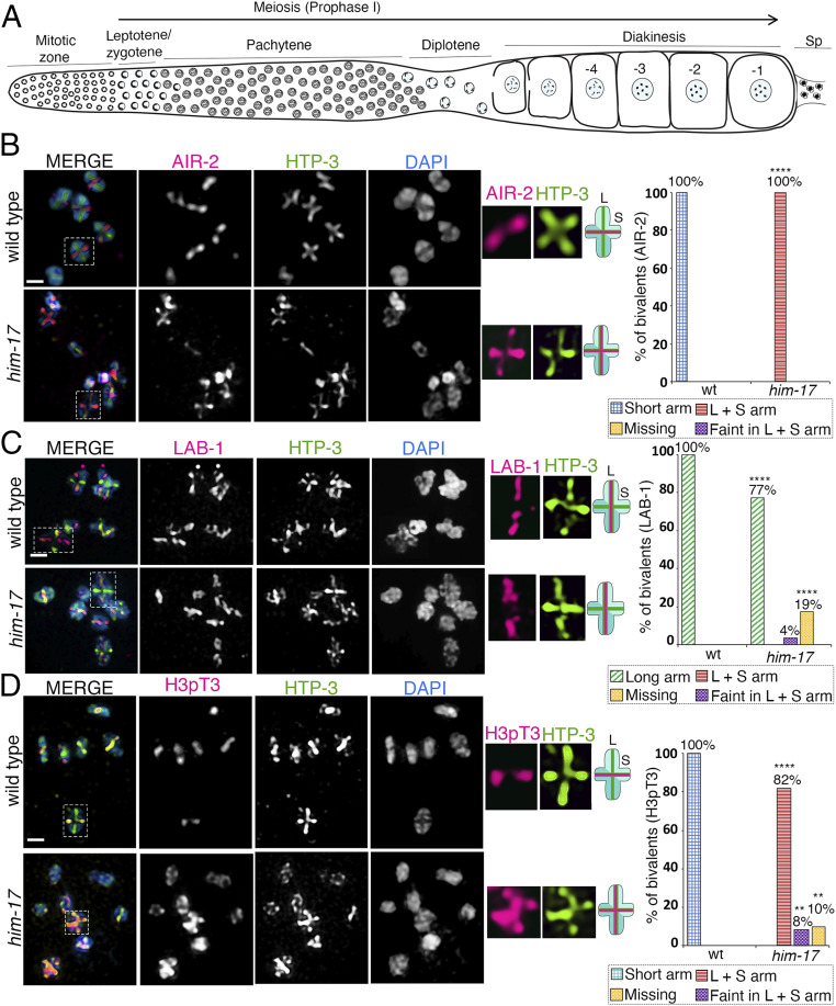
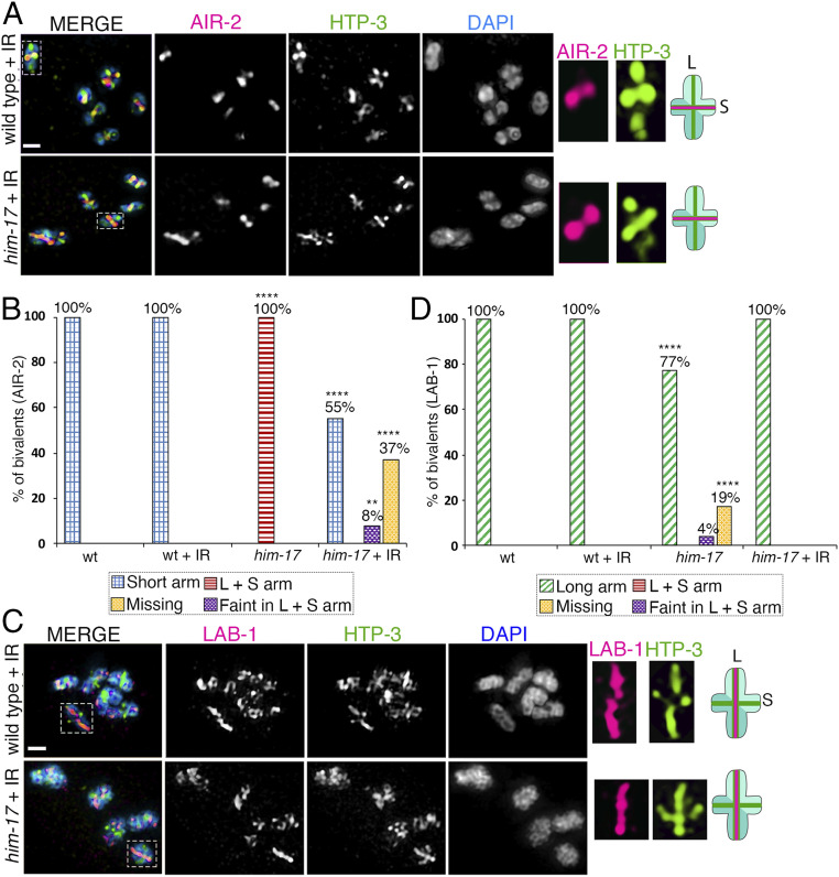
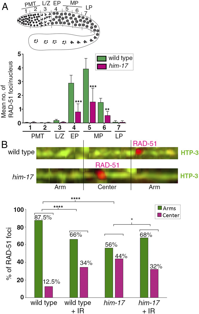
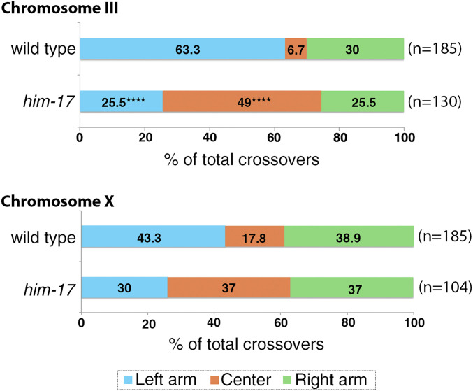
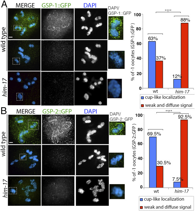
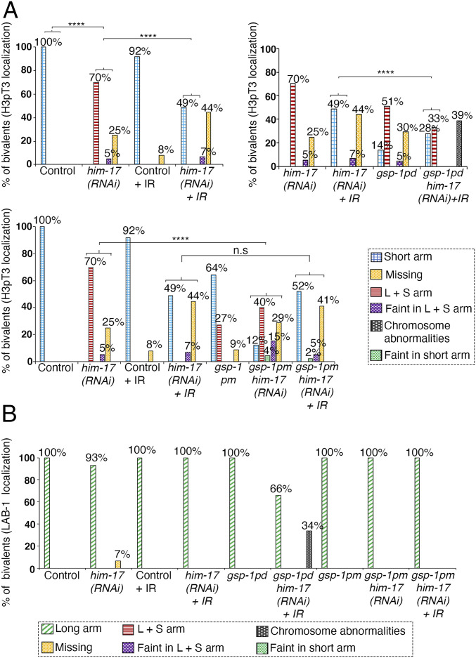
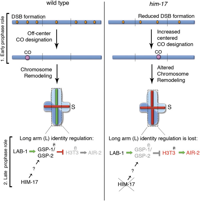
The position of recombination events established along chromosomes in early prophase I and the chromosome remodeling that takes place in late prophase I are intrinsically linked steps of meiosis that need to be tightly regulated to ensure accurate chromosome segregation and haploid gamete formation. Here, we show that RAD-51 foci, which form at the sites of programmed meiotic DNA double-strand breaks (DSBs), exhibit a biased distribution toward off-centered positions along the chromosomes in wild-type Caenorhabditis elegans, and we identify two meiotic roles for chromatin-associated protein HIM-17 that ensure normal chromosome remodeling in late prophase I. During early prophase I, HIM-17 regulates the distribution of DSB-dependent RAD-51 foci and crossovers on chromosomes, which is critical for the formation of distinct chromosome subdomains (short and long arms of the bivalents) later during chromosome remodeling. During late prophase I, HIM-17 promotes the normal expression and localization of protein phosphatases GSP-1/2 to the surface of the bivalent chromosomes and may promote GSP-1 phosphorylation, thereby antagonizing Aurora B kinase AIR-2 loading on the long arms and preventing premature loss of sister chromatid cohesion. We propose that HIM-17 plays distinct roles at different stages during meiotic progression that converge to promote normal chromosome remodeling and accurate chromosome segregation.

Keywords: DNA double-strand breaks; HIM-17; crossovers; late prophase I remodeling; meiosis.


Conflict of interest statement

The authors declare no competing interest.

Figures

Fig. 1.
Fig. 1.
HIM-17 regulates AIR-2 and H3pT3 localization on the short arm of the bivalents. High-magnification images of -1 oocytes at diakinesis in wild type and him-17 mutant are pictured. (A) Schematic representation of an adult C. elegans hermaphrodite gonad arm indicating nuclei in different stages of meiotic progression. The most proximal oocyte next to the spermatheca (Sp) is referred to as the -1 oocyte. (B) Costaining with anti–AIR-2 (magenta), anti–HTP-3 (green), and DAPI (blue) indicates that AIR-2 is not restricted to the short arm of the bivalents in him-17 mutants compared to wild type. Dashed boxes indicate the bivalents for which AIR-2 and HTP-3 localization are shown at higher magnification. (Middle) The chromosome configuration observed at this stage showing distinct short (S) and long (L) arms, and the localization of AIR-2 (magenta) and HTP-3 (green) for each genotype. (Right) Histogram indicates the percentage of bivalents at diakinesis with AIR-2 restricted to the short arm, mislocalized to both long and short arm, faint but in both long and short arms, or missing. A total of 102 and 97 bivalents were analyzed from the gonad arms of 18 and 112 animals for wild type and him-17, respectively, from three biological replicates. (C) Costaining with anti–LAB-1 (magenta), anti–HTP-3 (green), and DAPI (blue) indicates that a large number of bivalents in him-17 mutants exhibit LAB-1 localization restricted to the long arm as observed in wild type. (Right) Histogram indicates the percentage of bivalents at diakinesis with LAB-1 restricted to the long arm, long and short arm, faint but in both long and short arms, or missing on the chromosomes. A total of 90 and 96 bivalents from the gonad arms of 15 and 103 animals were analyzed for wild type and him-17, respectively, from three biological replicates. (D) Costaining with anti-H3pT3 (magenta), anti–HTP-3 (green), and DAPI (blue) indicates that H3pT3 signal is frequently observed on both long and short arms of the bivalents in him-17 mutants in contrast to the restricted localization only to the short arms observed in wild type. (Right) Histogram indicates the percentage of bivalents at diakinesis with H3pT3 localization restricted to the short arm, mislocalized to both long and short arms, faint but on both long and short arms, or missing on the chromosomes. A total of 99 and 72 bivalents from the gonad arms of 17 and 69 animals were analyzed for wild type and him-17, respectively, from three biological replicates. ****P < 0.0001, **P < 0.005 by the two-tailed Fisher’s exact test. (Scale bars, 2 μm.) For complete/additional statistical analysis, see Dataset S1.
Fig. 2.
Fig. 2.
AIR-2 and LAB-1 localization on bivalents in oocytes at diakinesis in wild type and him-17 mutants after exogenous DSB formation. High-magnification images of diakinesis stage oocytes in wild type and him-17 mutant after exogenous DSB formation by γ-IR (60 Gy). (A) Immunolocalization of AIR-2 (magenta), HTP-3 (green), and DAPI (blue) in -1 oocytes at diakinesis indicates that AIR-2 is restricted to the short arm of the bivalents in him-17 mutants, similar to wild type, after IR treatment. A total of 106 and 88 bivalents from the gonad arms of 20 and 16 animals were analyzed for wild type and him-17, respectively, from three biological replicates. Dashed boxes indicate the bivalents for which AIR-2 and HTP-3 localization are shown at higher magnification. (Middle) The chromosome configuration observed at this stage showing distinct short (S) and long (L) arms, and the localization of AIR-2 (magenta) and HTP-3 (green) for each genotype following IR treatment. (B) Histogram indicates the percentage of bivalents at diakinesis with AIR-2 restricted to the short arm, mislocalized to both long and short arms, faint but on both long and short arms, or missing from the chromosomes in wild type and him-17 mutants with and without IR treatment. (C) Immunolocalization of LAB-1 (magenta), HTP-3 (green), and DAPI (blue) in -1 oocytes at diakinesis. A total of 91 and 103 bivalents from the gonad arms of 16 and 18 animals were analyzed for wild type and him-17, respectively, from three biological replicates. (D) Histogram indicates the percentage of bivalents at diakinesis with LAB-1 restricted to the long arms, mislocalized to both long and short arms, faint but on both long and short arms, or missing from the chromosomes in wild type and him-17 mutants with and without IR. ****P < 0.0001, **P < 0.005 by the two-tailed Fisher’s exact test. (Scale bars, 2 μm.) For complete/additional statistical analysis, see Dataset S1.
Fig. 3.
Fig. 3.
Quantification of RAD-51 foci and distribution on the chromosomes in wild type and him-17 mutants. (A) Schematic representation of a C. elegans germline indicating the different zones scored for the number of RAD-51 foci/nucleus. (Bottom) Histogram depicts the mean number of RAD-51 foci/nucleus observed in different zones of him-17 mutant germlines compared to wild type. X-axis shows the position along the germline. PMT: premeiotic tip (germ cells in mitosis), L/Z: meiotic nuclei in leptotene/zygotene stage, EP: meiotic nuclei in early pachytene, MP: meiotic nuclei in midpachytene, and LP: meiotic nuclei in late pachytene. The number of nuclei scored per zone is indicated in Dataset S1. ***P < 0.001 and **P < 0.02 by the two-tailed Mann–Whitney test, 95% CI. (B, Top) Representative images of linearized chromosomes costained with anti–HTP-3 (green) to trace chromosome axes and anti–RAD-51 (magenta) to mark DSB repair sites. Linearized chromosomes from pachytene nuclei were divided into three equal portions referred to as arms and center. Only chromosomes with clear start and end points were scored using PRIISM software as in (25). (Bottom) Histogram indicates the distribution of RAD-51 foci on the center versus the arm regions of the chromosomes in wild type, wild type + IR, him-17, and him-17 + IR. A total of 115, 33, 113, and 36 chromosomes from pachytene stage nuclei from the gonad arms of 35, 6, 33, and 6 animals were analyzed for wild type, wild type + IR, him-17, and him-17 + IR, respectively. ****P < 0.0001, *P < 0.024 by the two-tailed Fisher’s exact test.
Fig. 4.
Fig. 4.
HIM-17 regulates CO distribution. An analysis of CO distribution on chromosomes III and X in wild type and him-17 mutants is shown. The positions on the chromosomes are indicated as left (blue), center (orange), and right (green). ****P < 0.0001 by two-tailed Fisher's exact test, 95% CI n = number of embryos scored.
Fig. 5.
Fig. 5.
GSP-1::GFP and GSP-2::GFP expression in wild type and him-17 mutant. (A) High-magnification images of diakinesis stage -1 oocytes from animals expressing GSP-1::GFP stained with anti-GFP (green) and DAPI (blue) in wild type and him-17 mutant backgrounds. Dashed boxes indicate the bivalents shown at higher magnification to the right for clearer visualization of either the presence or absence of GSP-1::GFP cup-like localization. (Right) Histogram indicates the percentage of -1 oocytes with cup-like localization of GSP-1::GFP on the chromosomes (blue) or a weak and diffuse signal not on the chromosomes (red) in wild type and him-17 mutant. A total of 30 and 33 -1 oocytes from 30 and 33 animals were scored for wild type and him-17, respectively, from three biological repeats. (B) High-magnification images of diakinesis stage -1 oocytes from animals expressing GSP-2::GFP stained with anti-GFP (green) and DAPI (blue) in wild type and him-17 mutants. Dashed boxes indicate the bivalents shown at higher magnification to the right for clearer visualization of either the presence or absence of GSP-2::GFP cup-like localization. (Right) Histogram indicates the percentage of -1 oocytes with cup-like localization of GSP-2::GFP on the chromosomes (blue) or weak and diffuse signal not on the chromosomes (red) in wild type and him-17 mutants. A total of 23 and 27 -1 oocytes from 23 and 27 animals were scored for wild type and him-17, respectively. ****P < 0.0001 two-tailed Fisher’s exact test. (Scale bars, 2 μm.)
Fig. 6.
Fig. 6.
Phosphorylation of GSP-1 at S2 partially rescues the mislocalization of H3pT3 observed in him-17 mutants. (A) Histograms indicate the percentage of bivalents at diakinesis with H3pT3 localization restricted to the short arm, mislocalized to both long and short arms, faint but on both long and short arms, faint but only on short arms, or missing on the chromosomes in the indicated genotypes. Chromosome abnormalities, consisting of aggregates and presence of DNA fragments, were observed in gsp-1pd him-17(RNAi) + IR. At least 100 bivalents were scored for each genotype from between 12 to 65 animals from three biological replicates. (B) Histogram indicates the percentage of bivalents at diakinesis with LAB-1 localization observed restricted to the long arm, mislocalized to both long and short arms, faint on both long and short arms, faint only on the short arm, or missing from chromosomes. Chromosome abnormalities, consisting of aggregates and DNA fragments, were observed in gsp-1pd him-17(RNAi) + IR. At least 72 bivalents were scored for each genotype from three biological replicates. ****P < 0.0001 two-tailed Fisher’s exact test. For complete/additional statistical analysis, see Dataset S1.
Fig. 7.
Fig. 7.
Model for how HIM-17 regulates DSB/CO distribution and late prophase I chromosome remodeling. HIM-17 acts in early prophase I by regulating DSB levels and DSB-dependent RAD-51 foci/CO distribution. In wild type, an excess number of DSBs are made, and a higher number of DSB-dependent RAD-51 foci is observed on the arms compared to the center region of the chromosomes in C. elegans. One DSB on the arm regions of the chromosomes is processed into a CO. This off-center CO position results in the production of an asymmetric bivalent with long (L) and short (S) arms. During late prophase I, recruitment of GSP-1/2 by LAB-1 and phosphorylation of GSP-1 at the S2 site, which requires HIM-17 function, prevents phosphorylation of H3T3 on the long arm, thereby preventing AIR-2 loading on the long arm. In contrast, in him-17 mutants, a reduced number of DSBs are made, and their distribution is altered such that now higher levels of DSB-dependent RAD-51 foci are detected at the center of the chromosomes. An increase in the levels of COs on the center of the chromosomes is observed, which potentially contributes to the short arm identity defect. In addition, expression, localization, and probably phosphorylation of GSP-1/2 is impaired, leading to phosphorylation of H3pT3 on the long arms even when LAB-1 is properly localized to the long arm of the bivalents. Therefore, HIM-17 functions at early and late prophase I are required to impede altered CO position and impaired GSP-1/2 activity to promote normal chromosome remodeling and subsequent regulated stepwise loss of SCC, leading to accurate chromosome segregation.

Similar articles

Cited by

References

    1. Keeney S., Giroux C. N., Kleckner N., Meiosis-specific DNA double-strand breaks are catalyzed by Spo11, a member of a widely conserved protein family. Cell 88, 375–384 (1997). - PubMed
    1. Keeney S., Lange J., Mohibullah N., Self-organization of meiotic recombination initiation: General principles and molecular pathways. Annu. Rev. Genet. 48, 187–214 (2014). - PMC - PubMed
    1. Bergerat A., et al. ., An atypical topoisomerase II from Archaea with implications for meiotic recombination. Nature 386, 414–417 (1997). - PubMed
    1. Dernburg A. F., et al. ., Meiotic recombination in C. elegans initiates by a conserved mechanism and is dispensable for homologous chromosome synapsis. Cell 94, 387–398 (1998). - PubMed
    1. Gray S., Cohen P. E., Control of meiotic crossovers: From double-strand break formation to designation. Annu. Rev. Genet. 50, 175–210 (2016). - PMC - PubMed

Publication types

MeSH terms