. 2021 Apr 21;11(1):8669.
doi: 10.1038/s41598-021-88159-x.

GLUT1 overexpression enhances glucose metabolism and promotes neonatal heart regeneration

Affiliations

GLUT1 overexpression enhances glucose metabolism and promotes neonatal heart regeneration

Viviana M Fajardo et al. Sci Rep. .

Abstract

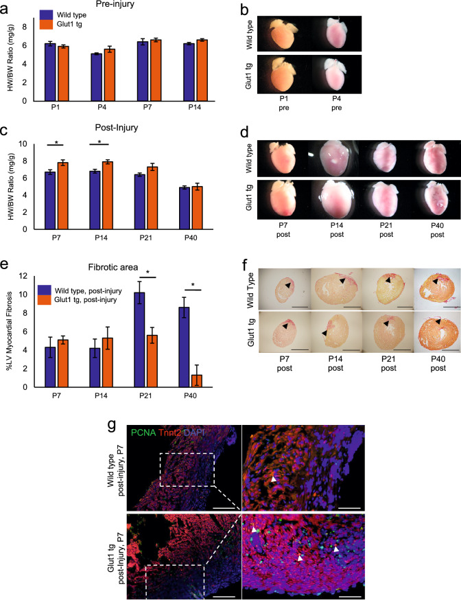
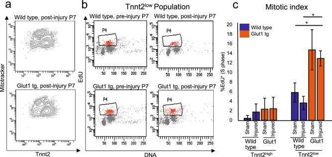
The mammalian heart switches its main metabolic substrate from glucose to fatty acids shortly after birth. This metabolic switch coincides with the loss of regenerative capacity in the heart. However, it is unknown whether glucose metabolism regulates heart regeneration. Here, we report that glucose metabolism is a determinant of regenerative capacity in the neonatal mammalian heart. Cardiac-specific overexpression of Glut1, the embryonic form of constitutively active glucose transporter, resulted in an increase in glucose uptake and concomitant accumulation of glycogen storage in postnatal heart. Upon cryoinjury, Glut1 transgenic hearts showed higher regenerative capacity with less fibrosis than non-transgenic control hearts. Interestingly, flow cytometry analysis revealed two distinct populations of ventricular cardiomyocytes: Tnnt2-high and Tnnt2-low cardiomyocytes, the latter of which showed significantly higher mitotic activity in response to high intracellular glucose in Glut1 transgenic hearts. Metabolic profiling shows that Glut1-transgenic hearts have a significant increase in the glucose metabolites including nucleotides upon injury. Inhibition of the nucleotide biosynthesis abrogated the regenerative advantage of high intra-cardiomyocyte glucose level, suggesting that the glucose enhances the cardiomyocyte regeneration through the supply of nucleotides. Our data suggest that the increase in glucose metabolism promotes cardiac regeneration in neonatal mouse heart.


Conflict of interest statement

The authors declare no competing interests.

Figures

Figure 1
Figure 1
Increase in intracellular glucose promotes cardiac regeneration in Glut1 transgenic heart. (a) Heart weight-body weight (HW/BW) ratio of Wild type and Glut1 transgenic (Glut1 tg) mice without injury. n = 3–7 for each group, p = n.s. (b) Representative images of hearts. (c) Heart weight-body weight (HW/BW) ratio of Wild type and Glut1 transgenic mouse post-injury. The hearts were cryoinjured at P1 and examined at P7, 14, 21 and 40. Note that the HW/BW is higher in Glut1 transgenic hearts at P7 and 14. n = 4–9 for each group, *p < 0.05. (d) Images of representative hearts post-injury. Note the ballooning of the heart at P14 in both wild type and Glut1 transgenic hearts. (e) %Fibrotic area measured by Image J capture of Picrosirius red stainings of the hearts. Hearts were cryoinjured at P1 and examined at P7, 14, 21 and 40. n = 5–8 for each group, *p < 0.05. (f) Representative images of Picrosirius red staining of wild type and Glut1 transgenic hearts. Arrowheads indicate fibrotic area. Scale bar = 200 µm. (g) PCNA staining of the sections from wild type and Glut1 transgenic hearts 7 days post-injury. Sections were stained with a cardiac marker (Tnnt2; Red), proliferation marker (PCNA; Green) and a nuclear marker (DAPI; Blue). Note that PCNA staining is more abundant in Glut1 transgenic heart. Arrowheads indicate PCNA positive cardiomyocytes. Scale bar = 50 µm and 20 µm respectively.
Figure 2
Figure 2
mRNA expression profile of Tnnt2high and Tnnt2low cardiomyocytes from Wild type and Glut1 transgenic hearts at P1. (a) Representative dot plot of flow cytometry analysis of P1 Wild type hearts for Tnnt2 (cardiac marker) and Mitotracker (mitochondrial contents). Note two populations of cardiomyocytes. (b) Flow cytometry analysis of P1 aMHC-Cre tg; R26+/YFP reporter and aMHC-hGLUT1 tg; aMHC-Cre tg; R26+/YFP reporter analysis for YFP (cardiac lineage) and Mitotracker (mitochondrial contents). Note two populations of cardiomyocytes. (c) Venn diagram of mRNA expressed in Tnnt2high and Tnnt2low cardiomyocytes from Wild type and Glut1 transgenic hearts. Bar graphs represent top three gene ontology (GO) term enriched in the genes uniquely expressed in each population. Although 4 populations are similar, G1 Tnnt2low cardiomyocytes are enriched for the genes associated with mitosis, cell cycle, and cell division. (d) GO analysis of the genes upregulated in Glut1 tg Tnnt2low vs Glut1 tg Tnnt2high. (e) Heatmap of expression level of representative cardiac, cell cycle, and mitochondrial genes differentially expressed in Glut1 tg Tnnt2low vs Glut1 tg Tnnt2high.
Figure 3
Figure 3
Tnnt2low cardiomyocytes are mitotically activated by the increase in intracellular glucose. (a) Representative flow cytometry profile of the ventricular cardiomyocytes from P7 Wild type and Glut1 transgenic hearts stained with Tnnt2 and Mitotracker. (b) Representative flow cytometry plot of EdU incorporation assay of Tnnt2low cardiomyocytes. Wild type and Glut1 transgenic mice were injected with EdU and the ventricular cardiomyocytes were isolated and analyzed by Tnnt2, Mitotracker, and EdU staining. (c) Mitotic index of Tnnt2high and Tnnt2low cardiomyocytes in Wild type and Glut1 transgenic heart with or without injury. n = 3–8. *p < 0.05. ***p < 0.001. Note that Tnn2low cardiomyocytes are more mitotic than Tnn2high cardiomyocytes and that the mitotic activity of Tnnt2low cardiomyocytes drastically increases in Glut1 transgenic background.
Figure 4
Figure 4
Metabolomics analysis of Glut1 transgenic hearts. (a) Metabolomics analysis of Glut1 transgenic hearts and wild type litters at P2 pre-injury and post-injury and P7 post-injury. At P2 stage, overall, the metabolic profile shows no drastic difference at the basal level, but glucose utility increases upon injury as represented by the increase in the metabolites in polyol pathway and glucose. At P7 stage, the metabolic profile showed a statistical differences of intermediates in the Pentose Phosphate Pathway. n = 3 wild type, n = 6 Glut1 tg hearts. *p < 0.05. **p < 0.01. (b) Histological analysis of glycogen storage of Glut1 transgenic hearts and its controls from wild type litters at P2, P4 and P7 by Periodic Acid Staining. Representative images of 3 samples in each group. Scale bar = 200 µm and 20 µm respectively. (c) Glycogen quantification by colorimetric assay kit at P2, P4 and P7 stage. n = 3 per group. *p < 0.05. **p < 0.01. Note the increased intracellular glycogen storage in Glut1 transgenic hearts.
Figure 5
Figure 5
Impact of hydroxyurea on the proliferation of Tnnt2low cardiomyocytes. (a) Representative contour plots (left) for Tnnt2 and Mitotracker and dot plots (right) for EdU and DNA content by flow cytometry analysis at P7. Wild type and Glut1 transgenic hearts were cryoinjured at P1 and treated with daily injection of HU or vehicle until the analysis at P7. (b) Quantification of %EdU positive cells in Tnnt2high and Tnnt2low cardiomyocytes in wild type and Glut1 transgenic hearts with or without HU treatment. Note that both Tnnt2high and Tnnt2low cardiomyocytes respond to HU treatment but that EdU incorporation of Tnnt2low cardiomyocytes was drastically decreased by HU treatment. n = 4. ***p < 0.01. (c) Quantification of the Tnnt2high and Tnnt2low fractions of cardiomyocytes in wild type and Glut1 transgenic hearts with or without HU treatment. While Tnnt2high or Tnnt2low fractions was not influenced by HU in wild type heart, HU treatment significantly decreased the percentage of Tnnt2low cardiomyocytes in Glut1 transgenic hearts. n = 4. **p < 0.01. ***p < 0.001.

References

    1. Porrello ER, et al. Transient regenerative potential of the neonatal mouse heart. Science (New York, N.Y.) 2011;331:1078–1080. doi: 10.1126/science.1200708. - DOI - PMC - PubMed
    1. Mollova M, et al. Cardiomyocyte proliferation contributes to heart growth in young humans. Proc. Natl. Acad. Sci. U.S.A. 2013;110:1446–1451. doi: 10.1073/pnas.1214608110. - DOI - PMC - PubMed
    1. Bergmann O, et al. Dynamics of cell generation and turnover in the human heart. Cell. 2015;161:1566–1575. doi: 10.1016/j.cell.2015.05.026. - DOI - PubMed
    1. Bergmann O, et al. Evidence for cardiomyocyte renewal in humans. Science (New York, N.Y.) 2009;324:98–102. doi: 10.1126/science.1164680. - DOI - PMC - PubMed
    1. Mahmoud AI, et al. Meis1 regulates postnatal cardiomyocyte cell cycle arrest. Nature. 2013;497:249–253. doi: 10.1038/nature12054. - DOI - PMC - PubMed

Publication types