

Effects of Intravitreous Aflibercept Injection in Pachychoroid Neovasculopathy: Comparison with Typical Neovascular Age-Related Macular Degeneration
- PMID: 33883872
- PMCID: PMC8054474
- DOI: 10.2147/OPTH.S285257
Effects of Intravitreous Aflibercept Injection in Pachychoroid Neovasculopathy: Comparison with Typical Neovascular Age-Related Macular Degeneration
Abstract
Purpose: To compare the 12-month efficacy of intravitreous aflibercept (IVA) injection between eyes with pachychoroid neovasculopathy and neovascular age-related macular degeneration (AMD).
Methods: Retrospective, comparative case series analysis. Twenty-seven eyes with pachychoroid neovasculopathy and sixty-three eyes with neovascular AMD. All patients received three initial monthly, followed by bimonthly, IVA injections.
Results: Twelve months after initial treatment, the mean best-corrected visual acuity (BCVA) had improved both in pachychoroid neovasculopathy (from 0.28 to 0.14 logMAR; P = 0.001) and neovascular AMD (from 0.40 to 0.29 logMAR; P < 0.001). Twelve months after initial treatment, eyes with pachychoroid neovasculopathy exhibited decreased mean central retinal thickness (CRT) and subfoveal choroidal thickness (both, P < 0.001) and presence of polyps (P = 0.039) and improved integrity of external limiting membrane (ELM) (P = 0.008) and ellipsoid zone band (P = 0.001). At the 12-month follow-up, 77% and 68% of eyes with pachychoroid neovasculopathy and neovascular AMD, respectively, exhibited dry macula (P = 0.30). Baseline CRT was correlated with 12-month BCVA in eyes with pachychoroid neovasculopathy (P = 0.02). In eyes with neovascular AMD, CRT (P = 0.005) and presence of intact ELM (P = 0.007) were significant predictors of 12-month BCVA.
Conclusion: Periodic IVA injection leads to anatomical and functional improvement in eyes with pachychoroid neovasculopathy and in eyes with neovascular AMD.
Keywords: aflibercept; age-related macular degeneration; choroidal thickness; pachychoroid neovasculopathy.
© 2021 Elfandi et al.
Conflict of interest statement
None of the authors has a proprietary interest in any of the products described herein. Dr Manabu Miyata reports personal fees from Santen Pharmaceutical, personal fees from HOYA, grants from Alcon Japan, grants from Novartis Pharma, outside the submitted work. Dr Hiroshi Tamura reports personal fees from Novartis, grants from Findex, and personal fees from Suntory, outside the submitted work. Dr Akio Oishi reports personal fees from Bayer and non-financial support from Tokai Optical, outside the submitted work. The authors report no other conflicts of interest in this work.
Figures
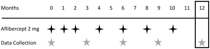
 Aflibercept injection (2 mg);
  clinical data collection;
  efficacy endpoint.

References
LinkOut - more resources
Full Text Sources
Other Literature Sources
Research Materials
