

Oral Tongue Cancer in a Patient with Fanconi Anemia: A Case Report and Literature Review
- PMID: 33883933
- PMCID: PMC8053604
- DOI: 10.2147/CMAR.S301582
Oral Tongue Cancer in a Patient with Fanconi Anemia: A Case Report and Literature Review
Abstract
Purpose: Fanconi anemia (FA) is a rare genetic disorder characterized by congenital anomalies, progressive bone marrow failure and high susceptibility to solid tumors, especially head and neck squamous cell carcinoma (HNSCC). Management of FA patients with head and neck cancer is a challenge due to increased risk of surgery, poor tolerance of chemotherapy, and severe myelotoxicity of radiotherapy.
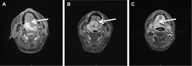
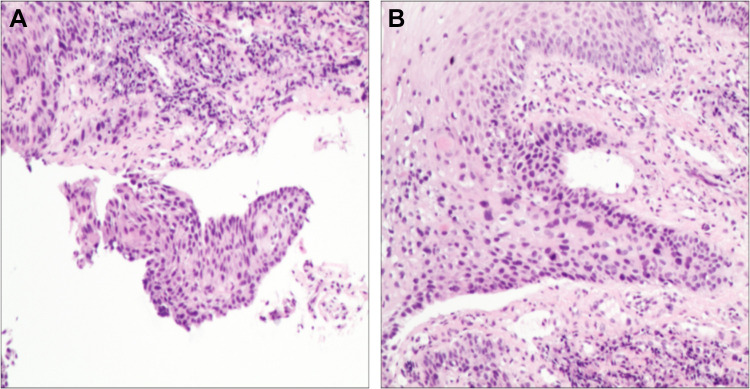
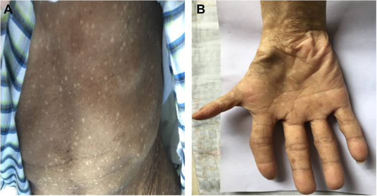
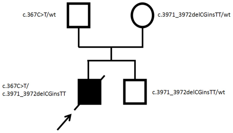
Patients and methods: We present a case of a 33-year-old man with carcinoma of oral tongue (T1N2M0), who experienced prolonged and profound bone marrow failure as a consequence of concurrent cisplatin/radiation. The young patient who developed HNSCC without risk factors, the myelotoxicity after exposure to platinum-based agent cisplatin and the further evaluation of phenotypic characteristics raised suspicion of FA. Whole exome sequencing performed for the patient and parents ultimately established the diagnosis of FA.
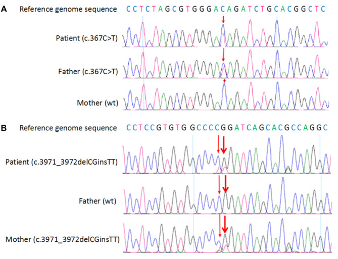
Results: Genetic testing in 23 FANC genes revealed two novel heterozygous mutations, c.367C>T and c.3971_3972delCGinsTT in FANCA gene of the patient, which were inherited from his father and mother, respectively. Radiotherapy with reduced dose has successfully alleviated the symptoms of tumor invasion and progression, and the radiation-related side effects were acceptable. Unfortunately, the patient eventually died of locoregional disease progression.
Conclusion: This case highlights the importance of considering the diagnosis of FA in young patients who develop HNSCC in the absence of risk factors, thus permitting more effective oncological treatment strategies and improved outcomes. In conclusion, any decision on different modalities of management in such patients should be based on a balance between locoregional control and therapeutic toxicity.
Keywords: cisplatin; fanconi anemia; head and neck squamous cell carcinoma; radiotherapy; toxicity.
© 2021 Deng et al.
Conflict of interest statement
The authors declare that the research was conducted in the absence of any commercial or financial relationships that could be construed as a potential conflict of interest.
Figures
Similar articles
-
Cisplatin-induced bone marrow failure in an adult patient with Fanconi anemia.J Oncol Pharm Pract. 2024 Oct;30(7):1274-1277. doi: 10.1177/10781552241268468. Epub 2024 Aug 2. J Oncol Pharm Pract. 2024. PMID: 39095039
-
Squamous cell carcinoma of the oral tongue in a patient with Fanconi anemia treated with radiotherapy and concurrent cetuximab: a case report and review of the literature.Head Neck. 2013 Oct;35(10):E292-8. doi: 10.1002/hed.23155. Epub 2012 Sep 10. Head Neck. 2013. PMID: 22965917 Review.
-
Strategies for early detection and detailed characterization of oral lesions and head and neck squamous cell carcinoma in Fanconi anemia patients.Cancer Lett. 2025 May 1;617:217529. doi: 10.1016/j.canlet.2025.217529. Epub 2025 Mar 5. Cancer Lett. 2025. PMID: 40054658 Review.
-
Assessing the spectrum of germline variation in Fanconi anemia genes among patients with head and neck carcinoma before age 50.Cancer. 2017 Oct 15;123(20):3943-3954. doi: 10.1002/cncr.30802. Epub 2017 Jul 5. Cancer. 2017. PMID: 28678401 Free PMC article.
-
Treatment of Fanconi Anemia-Associated Head and Neck Cancer: Opportunities to Improve Outcomes.Clin Cancer Res. 2021 Oct 1;27(19):5168-5187. doi: 10.1158/1078-0432.CCR-21-1259. Clin Cancer Res. 2021. PMID: 34045293 Free PMC article.
Cited by
-
Tongue cancer following hematopoietic cell transplantation for Fanconi anemia.Clin Oral Investig. 2022 Sep;26(9):5943-5952. doi: 10.1007/s00784-022-04554-2. Epub 2022 May 28. Clin Oral Investig. 2022. PMID: 35624384 Free PMC article.
-
Use of a Therapeutic Trial of Graduated Neoadjuvant Radiation Therapy for Locally Advanced Esophageal Cancer in a Patient With Fanconi Anemia.Adv Radiat Oncol. 2021 Sep 29;7(1):100810. doi: 10.1016/j.adro.2021.100810. eCollection 2022 Jan-Feb. Adv Radiat Oncol. 2021. PMID: 34765806 Free PMC article. No abstract available.
References
-
- Bhandari J, Thada PK, Puckett Y. Fanconi anemia. In: StatPearls. Treasure Island (FL): StatPearls Publishing LLC; 2020. - PubMed
-
- Furlong E, Carter T. Aplastic anaemia: current concepts in diagnosis and management. J Paediatr Child Health. 2020;56(7):1023–1028. - PubMed
-
- Kutler DI, Singh B, Satagopan J, et al. A 20-year perspective on the International Fanconi Anemia Registry (IFAR). Blood. 2003;101(4):1249–1256. - PubMed
-
- Meetei AR, Levitus M, Xue Y, et al. X-linked inheritance of Fanconi anemia complementation group B. Nat Genet. 2004;36(11):1219–1224. - PubMed
Publication types
LinkOut - more resources
Full Text Sources
Other Literature Sources
Miscellaneous
