

Binding of the SARS-CoV-2 Spike Protein to the Asialoglycoprotein Receptor on Human Primary Hepatocytes and Immortalized Hepatocyte-Like Cells by Confocal Analysis
- PMID: 33883951
- PMCID: PMC8055367
- DOI: 10.2147/HMER.S301979
Binding of the SARS-CoV-2 Spike Protein to the Asialoglycoprotein Receptor on Human Primary Hepatocytes and Immortalized Hepatocyte-Like Cells by Confocal Analysis
Abstract
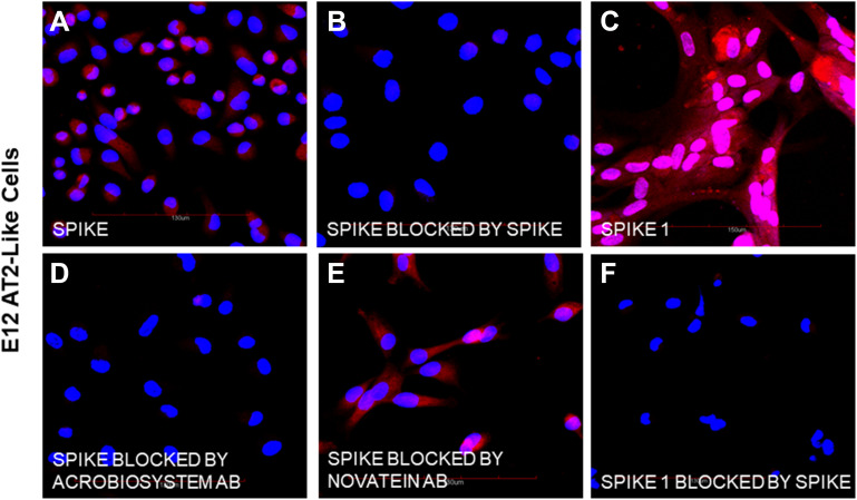
Background: The SARS-CoV-2 virus may have direct or indirect effects on other human organs beyond the respiratory system and including the liver, via binding of the spike protein. This study investigated the potential direct interactions with the liver by comparing the binding of SARS-CoV-2 spike proteins to human AT2-like cells, primary human hepatocytes and immortalized hepatocyte-like hybrid cells. Receptors with binding specificity for SARS-CoV-2 spike protein on AT2 cells and hepatocytes were identified.
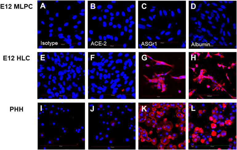
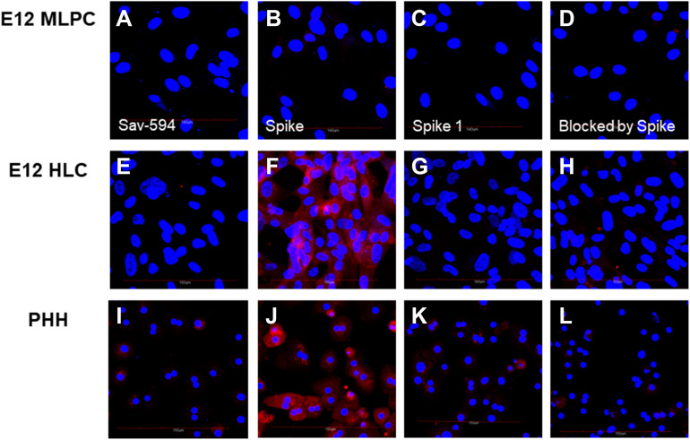
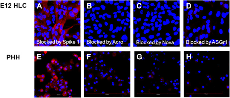
Methods: The specific binding of biotinylated spike and spike 1 proteins to undifferentiated human E12 MLPC (E12), E12 differentiated alveolar type 2 (AT2) cells, primary human hepatocytes (PHH) and E12 human hepatocyte-like hybrid cells (HLC) was studied by confocal microscopy. We investigated the expression of ACE-2, binding of biotinylated spike protein, biotinylated spike 1 and inhibition of binding by unlabeled spike protein, two neutralizing antibodies and an antibody directed against the hepatocyte asialoglycoprotein receptor 1 (ASGr1).
Results: E12 MLPC did not express ACE-2 and did not bind either of spike or spike 1 proteins. AT2-like cells expressed ACE-2 and bound both spike and spike 1. Both PHH and HLC did not express ACE-2 and did not bind spike 1 protein. However, both PHH and HLC actively bound the spike protein. Biotinylated spike protein binding was inhibited by unlabeled spike but not spike 1 protein on PHH and HLC. Two commercial neutralizing antibodies blocked the binding of the spike to PHH and HLC but only one blocked binding to AT2. An antibody to the hepatocyte ASGr1 blocked the binding of the spike protein to PHH and HLC.
Conclusion: The absence of ACE-2 receptors and inhibition of spike binding by an antibody to the ASGr1 on both PHH and HLC suggested that the spike protein interacts with the ASGr1. The differential antibody blocking of spike binding to AT2, PHH and HLC indicated that neutralizing activity of SARS-CoV-2 binding might involve additional mechanisms beyond RBD binding to ACE-2.
Keywords: AT2; E12 MLPC; SARS-CoV-2; asialoglycoprotein receptor; human hepatocytes; spike proteins.
© 2021 Collins and Steer.
Conflict of interest statement
Dr Daniel P Collins reports personal fees from BioE, LLC, during the conduct of the study. In addition, Dr Daniel P Collins has a patent “Composition for an in vitro culture medium to maintain and expand stem cell-derived hepatocyte-like cells” pending, as well as, a patent “Methods to develop immortalized hybrid hepatocyte-like cells”, also pending. The authors report no other conflicts of interest in this work.
Figures
Similar articles
-
Differentiation of immortalized human multi-lineage progenitor to alveolar type 2-like cells: angiotensin-converting enzyme 2 expression and binding of severe acute respiratory syndrome coronavirus 2 spike and spike 1 proteins.Cytotherapy. 2021 Dec;23(12):1064-1073. doi: 10.1016/j.jcyt.2021.07.017. Epub 2021 Aug 17. Cytotherapy. 2021. PMID: 34551876 Free PMC article.
-
Analysis of SARS-CoV-2 Variant-Specific Serum Antibody Post-Vaccination Utilizing Immortalized Human Hepatocyte-Like Cells (HLC) to Assess Development of Immunity.Hepat Med. 2023 Dec 5;15:221-231. doi: 10.2147/HMER.S431327. eCollection 2023. Hepat Med. 2023. PMID: 38078048 Free PMC article.
-
Identification of an FXR-modulated liver-intestine hybrid state in iPSC-derived hepatocyte-like cells.J Hepatol. 2022 Nov;77(5):1386-1398. doi: 10.1016/j.jhep.2022.07.009. Epub 2022 Jul 19. J Hepatol. 2022. PMID: 35863491
-
Comprehensive characterization of the antibody responses to SARS-CoV-2 Spike protein finds additional vaccine-induced epitopes beyond those for mild infection.Elife. 2022 Jan 24;11:e73490. doi: 10.7554/eLife.73490. Elife. 2022. PMID: 35072628 Free PMC article.
-
Development of immortalized human hepatocyte-like hybrid cells by fusion of multi-lineage progenitor cells with primary hepatocytes.PLoS One. 2020 Jun 4;15(6):e0234002. doi: 10.1371/journal.pone.0234002. eCollection 2020. PLoS One. 2020. PMID: 32497071 Free PMC article.
Cited by
-
Differentiation of immortalized human multi-lineage progenitor to alveolar type 2-like cells: angiotensin-converting enzyme 2 expression and binding of severe acute respiratory syndrome coronavirus 2 spike and spike 1 proteins.Cytotherapy. 2021 Dec;23(12):1064-1073. doi: 10.1016/j.jcyt.2021.07.017. Epub 2021 Aug 17. Cytotherapy. 2021. PMID: 34551876 Free PMC article.
-
Protein structure-based in-silico approaches to drug discovery: Guide to COVID-19 therapeutics.Mol Aspects Med. 2023 Jun;91:101151. doi: 10.1016/j.mam.2022.101151. Epub 2022 Oct 28. Mol Aspects Med. 2023. PMID: 36371228 Free PMC article. Review.
-
SARS-CoV-2-Specific Immune Response and the Pathogenesis of COVID-19.Int J Mol Sci. 2022 Feb 2;23(3):1716. doi: 10.3390/ijms23031716. Int J Mol Sci. 2022. PMID: 35163638 Free PMC article. Review.
-
Expression of plasma IFN signaling-related miRNAs during acute SARS-CoV-2 infection and its association with RBD-IgG antibody response.Virol J. 2021 Dec 7;18(1):244. doi: 10.1186/s12985-021-01717-7. Virol J. 2021. PMID: 34876159 Free PMC article.
-
Non-alcoholic fatty liver disease and COVID-19: Harmless companions or disease intensifier?World J Gastroenterol. 2023 Jan 14;29(2):367-377. doi: 10.3748/wjg.v29.i2.367. World J Gastroenterol. 2023. PMID: 36687116 Free PMC article. Review.
References
LinkOut - more resources
Full Text Sources
Other Literature Sources
Miscellaneous
