

Microcirculatory disturbance in acute liver injury
- PMID: 33884034
- PMCID: PMC8056117
- DOI: 10.3892/etm.2021.10028
Microcirculatory disturbance in acute liver injury
Abstract
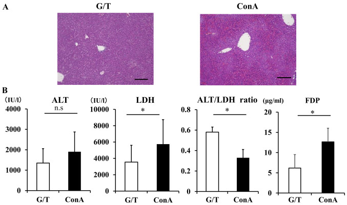
Microcirculatory disturbance is thought to be involved in the pathogenesis of acute liver injury (ALI). The current study examined the pathophysiologic role of hepatic microcirculatory disturbance in patients with ALI and in mouse models of ALI. Using serum aminotransferase (ALT)/lactate dehydrogenase (LDH) ratio as a hypoxic marker, 279 patients with ALI were classified into the low ALT/LDH ratio (ALT/LDH ≤1.5) and high ALT/LDH ratio group (ALT/LDH >1.5). In the low ALT/LDH ratio group, serum ALT, LDH, fibrinogen degradation products and prothrombin time-international normalized ratio were increased relative to the high ALT/LDH ratio group. Histologically, hepatic expression of tissue factor (TF) and hypoxia-related proteins was enhanced in the low ALT/LDH ratio group, and this was accompanied by sinusoidal fibrin deposition. Sinusoidal hypercoagulation and intrahepatic hypoxia was also analyzed in two different mouse models of ALI; Concanavalin A (ConA) mice and Galactosamine/tumor necrosis factor (TNF)-α (G/T) mice. Serum ALT/LDH ratio in ConA mice was significantly lower compared with G/T mice. Pimonidazole staining revealed the upregulation of hypoxia-related proteins in ConA mice. Recombinant human soluble thrombomodulin improved liver damage in ConA mice in association with reduced sinusoidal hypercoagulation and intrahepatic hypoxia. The present study provides evidence that serum ALT/LDH ratio aids in the identification of patients with ALI and intrahepatic hypoxia as a result of microcirculatory disturbance. The results facilitate the improved understanding of the pathogenesis of ALI, thereby offering a novel therapeutic strategy against ALI, which arises from sinusoidal hypercoagulation.
Keywords: acute liver failure; intrahepatic hypoxia; microcirculatory disturbance; serum aminotransferase/lactate dehydrogenase ratio; sinusoidal hypercoagulation.
Copyright: © Kuwano et al.
Conflict of interest statement
The authors declare that they have no competing interests.
Figures


Similar articles
-
Microcirculatory disturbance in acute liver injury is triggered by IFNγ-CD40 axis.J Inflamm (Lond). 2024 Jun 21;21(1):23. doi: 10.1186/s12950-024-00387-w. J Inflamm (Lond). 2024. PMID: 38907339 Free PMC article.
-
A new parameter using serum lactate dehydrogenase and alanine aminotransferase level is useful for predicting the prognosis of patients at an early stage of acute liver injury: a retrospective study.Comp Hepatol. 2008 Aug 14;7:6. doi: 10.1186/1476-5926-7-6. Comp Hepatol. 2008. PMID: 18700988 Free PMC article.
-
Ischaemic Markers in Acute Hepatic Injury.J Clin Diagn Res. 2016 Apr;10(4):BC17-20. doi: 10.7860/JCDR/2016/16699.7680. Epub 2016 Apr 1. J Clin Diagn Res. 2016. PMID: 27190791 Free PMC article.
-
Cooperation of liver cells in health and disease.Adv Anat Embryol Cell Biol. 2001;161:III-XIII, 1-151. doi: 10.1007/978-3-642-56553-3. Adv Anat Embryol Cell Biol. 2001. PMID: 11729749 Review.
-
Hepatic microcirculatory disturbance in liver diseases: intervention with traditional Chinese medicine.Front Pharmacol. 2024 Jul 23;15:1399598. doi: 10.3389/fphar.2024.1399598. eCollection 2024. Front Pharmacol. 2024. PMID: 39108760 Free PMC article. Review.
Cited by
-
Cardiac Hepatopathy: New Perspectives on Old Problems through a Prism of Endogenous Metabolic Regulations by Hepatokines.Antioxidants (Basel). 2023 Feb 17;12(2):516. doi: 10.3390/antiox12020516. Antioxidants (Basel). 2023. PMID: 36830074 Free PMC article. Review.
-
Priming, Triggering, Adaptation and Senescence (PTAS): A Hypothesis for a Common Damage Mechanism of Steatohepatitis.Int J Mol Sci. 2021 Nov 21;22(22):12545. doi: 10.3390/ijms222212545. Int J Mol Sci. 2021. PMID: 34830427 Free PMC article.
-
Laboratory scoring system to predict hepatic indocyanine green clearance ability during fluorescence imaging-guided laparoscopic hepatectomy.World J Gastrointest Surg. 2023 Jul 27;15(7):1442-1453. doi: 10.4240/wjgs.v15.i7.1442. World J Gastrointest Surg. 2023. PMID: 37555108 Free PMC article. Clinical Trial.
-
Mitochondrial Lipid Peroxidation and Microsomal Drug-metabolizing Enzyme Activity of Rat Hepatotoxicity under Heavy Metals from Slag Waste Exposure.Cell Biochem Biophys. 2023 Jun;81(2):285-298. doi: 10.1007/s12013-023-01134-3. Epub 2023 Jun 3. Cell Biochem Biophys. 2023. PMID: 37268808
-
Microcirculatory disturbance in acute liver injury is triggered by IFNγ-CD40 axis.J Inflamm (Lond). 2024 Jun 21;21(1):23. doi: 10.1186/s12950-024-00387-w. J Inflamm (Lond). 2024. PMID: 38907339 Free PMC article.
References
LinkOut - more resources
Full Text Sources
Other Literature Sources
Miscellaneous