

E. coli RNase I exhibits a strong Ca2+-dependent inherent double-stranded RNase activity
- PMID: 33885787
- PMCID: PMC8136782
- DOI: 10.1093/nar/gkab284
E. coli RNase I exhibits a strong Ca2+-dependent inherent double-stranded RNase activity
Abstract
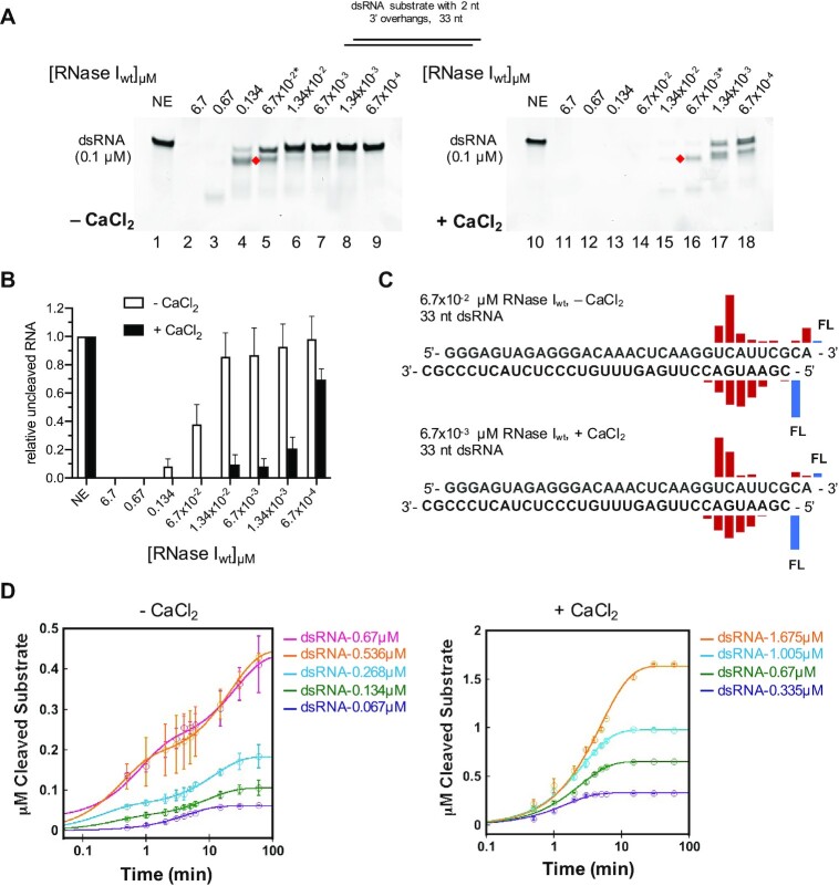
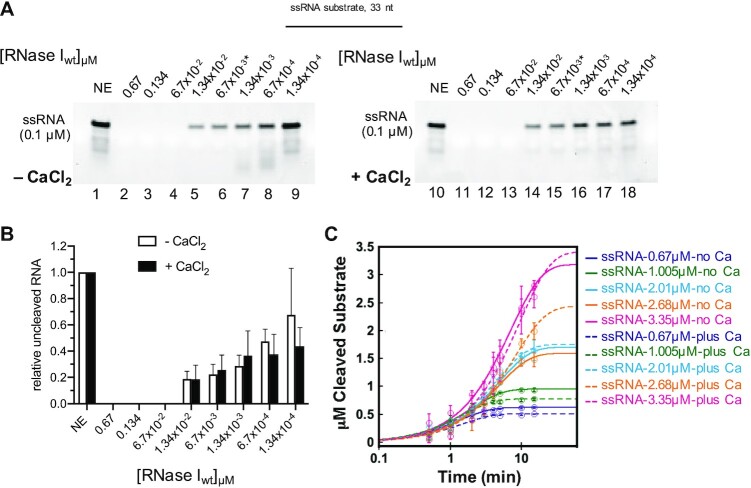
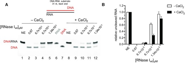
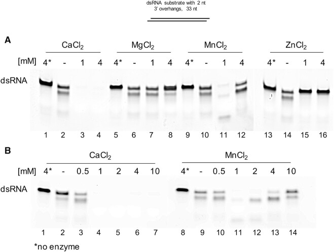
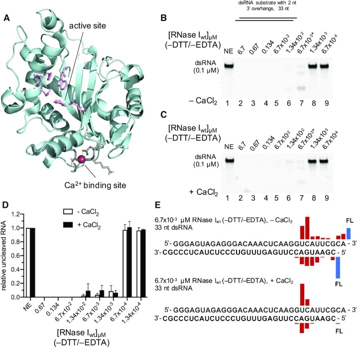
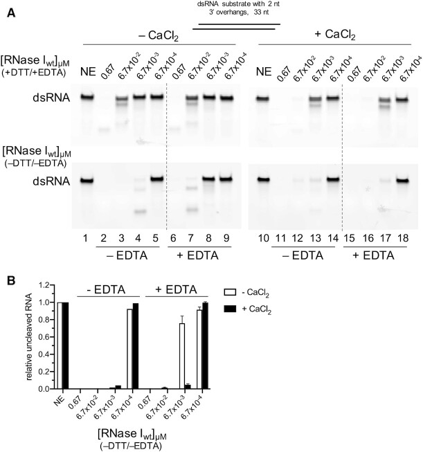
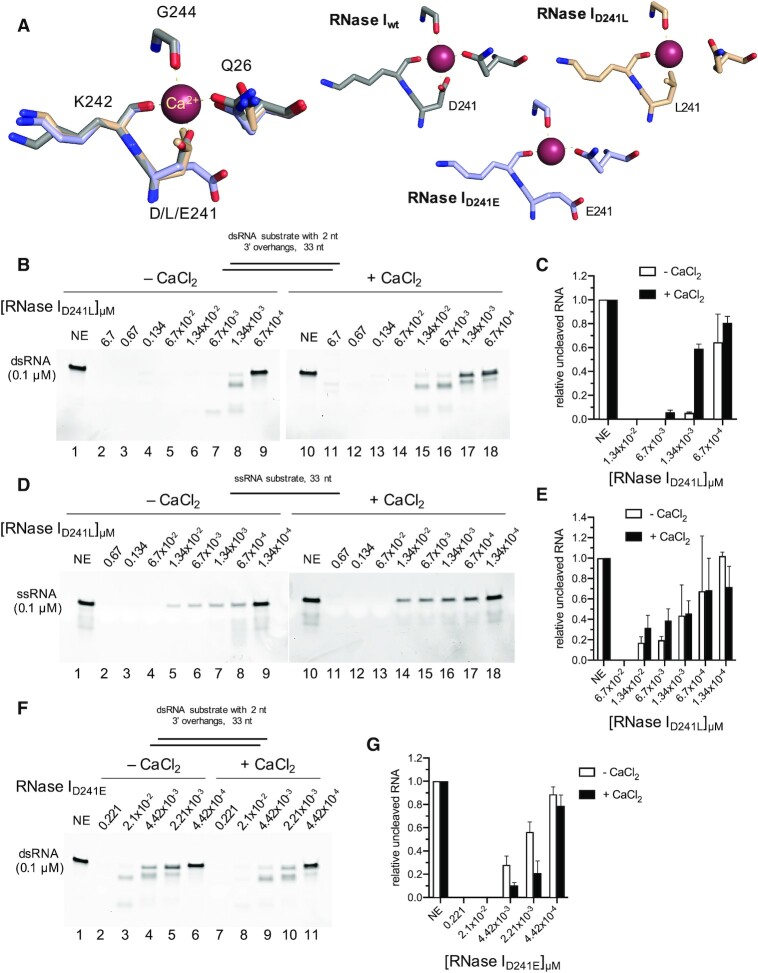
Since its initial characterization, Escherichia coli RNase I has been described as a single-strand specific RNA endonuclease that cleaves its substrate in a largely sequence independent manner. Here, we describe a strong calcium (Ca2+)-dependent activity of RNase I on double-stranded RNA (dsRNA), and a Ca2+-dependent novel hybridase activity, digesting the RNA strand in a DNA:RNA hybrid. Surprisingly, Ca2+ does not affect the activity of RNase I on single stranded RNA (ssRNA), suggesting a specific role for Ca2+ in the modulation of RNase I activity. Mutation of a previously overlooked Ca2+ binding site on RNase I resulted in a gain-of-function enzyme that is highly active on dsRNA and could no longer be stimulated by the metal. In summary, our data imply that native RNase I contains a bound Ca2+, allowing it to target both single- and double-stranded RNAs, thus having a broader substrate specificity than originally proposed for this traditional enzyme. In addition, the finding that the dsRNase activity, and not the ssRNase activity, is associated with the Ca2+-dependency of RNase I may be useful as a tool in applied molecular biology.
© The Author(s) 2021. Published by Oxford University Press on behalf of Nucleic Acids Research.
Figures
Similar articles
-
Double-stranded RNA-dependent RNase activity associated with human immunodeficiency virus type 1 reverse transcriptase.Proc Natl Acad Sci U S A. 1992 Feb 1;89(3):927-31. doi: 10.1073/pnas.89.3.927. Proc Natl Acad Sci U S A. 1992. PMID: 1371014 Free PMC article.
-
Intrinsic double-stranded-RNA processing activity of Escherichia coli ribonuclease III lacking the dsRNA-binding domain.Biochemistry. 2001 Dec 11;40(49):14976-84. doi: 10.1021/bi011570u. Biochemistry. 2001. PMID: 11732918
-
Genetic uncoupling of the dsRNA-binding and RNA cleavage activities of the Escherichia coli endoribonuclease RNase III--the effect of dsRNA binding on gene expression.Mol Microbiol. 1998 May;28(3):629-40. doi: 10.1046/j.1365-2958.1998.00828.x. Mol Microbiol. 1998. PMID: 9632264
-
The RNase III family: a conserved structure and expanding functions in eukaryotic dsRNA metabolism.Curr Issues Mol Biol. 2001 Oct;3(4):71-8. Curr Issues Mol Biol. 2001. PMID: 11719970 Review.
-
Dissecting nucleases into their structural and functional domains. Mapping the RNA-binding surface of RNase III by NMR.Methods Mol Biol. 2001;160:237-48. doi: 10.1385/1-59259-233-3:237. Methods Mol Biol. 2001. PMID: 11265286 Review. No abstract available.
Cited by
-
Incorporation of noncanonical base Z yields modified mRNA with minimal immunogenicity and improved translational capacity in mammalian cells.iScience. 2023 Aug 26;26(10):107739. doi: 10.1016/j.isci.2023.107739. eCollection 2023 Oct 20. iScience. 2023. PMID: 37720088 Free PMC article.
-
Preanalytical Impact of Incomplete K2EDTA Blood Tube Filling in Molecular Biology Testing.Diagnostics (Basel). 2024 Sep 2;14(17):1934. doi: 10.3390/diagnostics14171934. Diagnostics (Basel). 2024. PMID: 39272719 Free PMC article.
-
Insights into the metabolism, signaling, and physiological effects of 2',3'-cyclic nucleotide monophosphates in bacteria.Crit Rev Biochem Mol Biol. 2023 Dec;58(2-6):118-131. doi: 10.1080/10409238.2023.2290473. Epub 2024 Feb 2. Crit Rev Biochem Mol Biol. 2023. PMID: 38064689 Free PMC article. Review.
-
Methods for the Cost-Effective Production of Bacteria-Derived Double-Stranded RNA for in vitro Knockdown Studies.Front Physiol. 2022 Apr 13;13:836106. doi: 10.3389/fphys.2022.836106. eCollection 2022. Front Physiol. 2022. PMID: 35492592 Free PMC article.
-
SARS-CoV2 Nsp1 is a metal-dependent DNA and RNA endonuclease.Biometals. 2024 Oct;37(5):1127-1146. doi: 10.1007/s10534-024-00596-z. Epub 2024 Mar 28. Biometals. 2024. PMID: 38538957 Free PMC article.
References
-
- Deshpande R.A., Shankar V.. Ribonucleases from T2 family. Crit. Rev. Microbiol. 2002; 28:79–122. - PubMed
-
- Irie M. Structure-function relationships of acid ribonucleases: lysosomal, vacuolar, and periplasmic enzymes. Pharmacol. Ther. 1999; 81:77–89. - PubMed
-
- Aravind L., Koonin E.V.. A natural classification of ribonucleases. Methods Enzymol. 2001; 341:3–28. - PubMed
-
- Spahr P.F., Hollingworth B.R.. Purification and mechanism of action of ribonuclease from Escherichia coli ribosomes. J. Biol. Chem. 1961; 236:823–831.
Publication types
MeSH terms
Substances
LinkOut - more resources
Full Text Sources
Other Literature Sources
Miscellaneous
