. 2021 May 21;49(9):5216-5229.
doi: 10.1093/nar/gkab277.

A nucleotide-sensing endonuclease from the Gabija bacterial defense system

Affiliations

A nucleotide-sensing endonuclease from the Gabija bacterial defense system

Rui Cheng et al. Nucleic Acids Res. .

Abstract

The arms race between bacteria and phages has led to the development of exquisite bacterial defense systems including a number of uncharacterized systems distinct from the well-known restriction-modification and CRISPR/Cas systems. Here, we report functional analyses of the GajA protein from the newly predicted Gabija system. The GajA protein is revealed as a sequence-specific DNA nicking endonuclease unique in that its activity is strictly regulated by nucleotide concentration. NTP and dNTP at physiological concentrations can fully inhibit the robust DNA cleavage activity of GajA. Interestingly, the nucleotide inhibition is mediated by an ATPase-like domain, which usually hydrolyzes ATP to stimulate the DNA cleavage when associated with other nucleases. These features suggest a mechanism of the Gabija defense in which an endonuclease activity is suppressed under normal conditions, while it is activated by the depletion of NTP and dNTP upon the replication and transcription of invading phages. This work highlights a concise strategy to utilize a DNA nicking endonuclease for phage resistance via nucleotide regulation.


Figures

Figure 1.
Figure 1.
Purified GajA as an endonuclease. (A) Domain architecture of GajA protein. (B) SDS-PAGE gel showing purified GajA (69 kDa including an N-terminal His-tag). (C) Cleavage of linear λDNA by GajA. (D) GajA nuclease activity is dependent on metal ions. Reaction mixtures containing 20 mM Tris–HCl pH 9, 0.1 mg/ml BSA, 20 nM λ955 DNA, 200 nM GajA and 5 mM metal ions (MgCl2, MnCl2, CaCl2, ZnCl2, CoCl2 or NiCl2 as shown on top of the gel) were incubated at 37°C for 5 min. Gel bands corresponding to the 955-bp λ955 DNA substrate and 583-bp and 372-bp DNA products resulting from specific endonuclease cleavage are annotated. Reactions with no metal ions added (-) and 5 mM EDTA were included as controls. (E) Recovery of the two DNA fragments (λ372 and λ583) from GajA endonuclease cleavage for cloning and sequencing. (F) The GajA cleavage site and pattern were determined based on DNA sequencing. The λ372 and λ583 fragments were inserted into T-plasmids and the regions near the cleavage site were respectively sequenced. Red sequences are the terminal sequences of each fragment derived from the sequencing results (bottom), and the dotted red lines demonstrate the cleavage site.
Figure 2.
Figure 2.
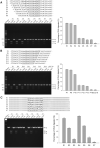
Characterization of the full optimal recognition sequence of GajA. The preliminary recognition sequence of GajA was shortened one nucleotide at a time from the left end (A) or right end (B), and DNA fragments containing the resulting sequences were PCR-amplified to be used as substrates for GajA. (C) Cleavage efficiency (measured as a reduction of initial DNA substrates) of GajA on DNA oligos with base-switching in the palindromic region. The synthetic DNA substrates were prepared by mixing equimolar amounts of complementary 56-nt oligonucleotides. The DNA substrate (800 nM) containing one GajA recognition site was digested by GajA (400 nM), and results were analyzed by 10% PAGE. DNA digestion was measured using ImageJ software as described in the Materials and Methods. All graphs represent the average of three independent trials with error bars representing the standard error of the mean.
Figure 3.
Figure 3.
Turnover and efficiency of GajA. (A) Cleavage efficiency of GajA at different molar ratios of enzyme to substrate. The synthetic DNA oligo S1 (0.8 μM) was incubated with GajA (0.4 μM) and the reaction time was set as 5, 10, or 20 min, respectively. (B) GajA exhibits rapid DNA cleavage activity. λ955 DNA was used as the substrate. Over 60% of the DNA substrate (20 nM) was digested by GajA (200 nM) after 30 s, and over 96% was digested after 120 s. Reactions were performed in a final volume of 10 μl in the optimal reaction buffer at 37°C and then stopped by addition of 2 μl of 6× loading dye containing 20 mM EDTA. Samples were analyzed via native agarose gel electrophoresis. All graphs represent the average of three independent trials with error bars representing the standard error of the mean.
Figure 4.
Figure 4.
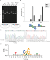
GajA is a site-specific nicking enzyme. (A) GajA cleavage patterns on various plasmids. The P1 plasmid contains the complete GajA restriction sequence consisting of two overlapping minimum recognition sequences, P13 contains one minimum recognition sequence, and pUC19 without the GajA recognition sequence was used as a control. The sequence of the colored DNA region in P1 is 5′-AATAACCCGGATATT-3′ and that in P13 is 5′-AATAACCCGG-3′. ‘N’, ‘L’, and ‘S’ denote the positions of gel bands corresponding to ‘nicked’, ‘linearized’, and ‘supercoiled’ DNA, respectively. In the plasmid diagram, the core GC-rich region of the GajA recognition sequence is shown in red and the AT-rich wing region in blue. (B) Quantification of products of GajA cleavage on plasmids P1 and P13. Proportions of nicked, linearized, and supercoiled DNA after GajA treatment were compared. The bottom diagram depicts the two action modes of GajA endonuclease. (C) Nicking site of nicked P13 plasmid after incubation with GajA. The nicked DNA of P13 plasmid after incubation with GajA was recycled separately and subjected to DNA sequencing. The overlapping double peak indicates the nicked site (denoted by the arrow). The forward sequencing result exhibited an additional peak corresponding to ‘A’, while the reverse sequencing was normal. (D) The nicking sites of GajA in six fragments of T7 genomic DNA compiled by the WebLogo server. The overall height of each stack indicates the sequence conservation at that position (measured in bits), and the height of symbols within the stack reflects the relative frequency of the corresponding base at that position. The arrow indicates nicking site.
Figure 5.
Figure 5.
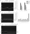
The endonuclease activity of GajA is inhibited by nucleotides. (A) Representative gel and quantification of GajA endonuclease activity on λ955 DNA in the presence of increasing amounts of ATP and AMP-PNP. (B) Effect of NTP, dNTP, ADP, and AMP on GajA endonuclease activity. (C) Effect of NDP, NMP, dNMP and nucleosides on GajA endonuclease activity. All reactions contained 20 nM λ955 DNA and 200 nM GajA and were incubated at 37°C for 5 min. Lanes labeled with dashes indicate no nucleotide addition. Initial DNA digested was quantified using ImageJ software. Bar graphs represent the average of three independent experiments with error bars representing the standard error of the mean.
Figure 6.
Figure 6.
Investigation of GajA functional domains by site-specific mutagenesis. (A) SDS-PAGE analysis of purified wild-type (WT) GajA, GajA mutants and the C-terminal polypeptide (CTR) of GajA. (B) Endonuclease activity of the proteins in (A). (C) The effect of K35A or H320A mutations on the ATP inhibition of GajA activity. H320A but not K35A mutation partially relieved the inhibition of ATP on GajA activity. For (B) and (C), 125 ng of λ955 DNA (20 nM) was incubated with 0.2 μM GajA in a final volume of 10 μl in the optimal reaction buffer with or without 0.5 mM ATP. Reactions were performed at 37°C for 5 min and then stopped by addition of 2 μl of 6× loading dye containing 20 mM EDTA. Samples were analyzed via native agarose gel electrophoresis. Bar graphs represent the average of three independent experiments with error bars representing the standard error of the mean.
Figure 7.
Figure 7.
Schematic showing the proposed mechanism of Gabija anti-phage bacterial defense. The DNA nicking of GajA on the genomic DNA of bacteriophage T7 (A) or E. coli B (ATCC® 11303™) (B) in the absence or presence of 0.5 mM ATP. (C) Under normal conditions, GajA endonuclease activity is fully inhibited by nucleotides at physiological concentrations in bacteria. The ATPase-like domain of GajA senses and binds NTP and dNTP to allosterically regulate the TOPRIM domain. (D) During phage invasion, active phage transcription and DNA replication deplete cellular NTP and dNTP. When NTP and dNTP concentrations decrease to a certain degree, the loss of nucleotide binding of the GajA ATPase-like domain activates the TOPRIM domain. The latter, in turn, mediates phage DNA cleavage and may also mediate destruction of bacterial genomic DNA for abortive infection. GajB may contribute to GajA activation or facilitate GajA cleavage, which is under investigation and not shown in this model.

References

    1. Dy R.L., Richter C., Salmond G.P.C., Fineran P.C.. Remarkable mechanisms in microbes to resist phage infections. Annu. Rev. Virol. 2014; 1:307–331. - PubMed
    1. Labrie S.J., Samson J.E., Moineau S.. Bacteriophage resistance mechanisms. Nat. Rev. Microbiol. 2010; 8:317–327. - PubMed
    1. Stern A., Sorek R.. The phage-host arms race: Shaping the evolution of microbes. Bioessays. 2011; 33:43–51. - PMC - PubMed
    1. Barrangou R., Fremaux C., Deveau H., Richards M., Boyaval P., Moineau R., S. D.A., Horvath P.. CRISPR provides acquired resistance against viruses in prokaryotes. Science. 2007; 315:1709–1712. - PubMed
    1. Tock M.R., Dryden D.T.F.. The biology of restriction and anti-restriction. Curr. Opin. Microbiol. 2005; 8:466–472. - PubMed

Publication types

MeSH terms

LinkOut - more resources