. 2021 Jun 2;109(11):1825-1835.e5.
doi: 10.1016/j.neuron.2021.03.038. Epub 2021 Apr 21.

Translation of GGC repeat expansions into a toxic polyglycine protein in NIID defines a novel class of human genetic disorders: The polyG diseases

Affiliations

Translation of GGC repeat expansions into a toxic polyglycine protein in NIID defines a novel class of human genetic disorders: The polyG diseases

Manon Boivin et al. Neuron. .

Abstract

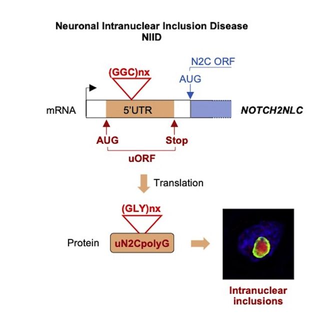
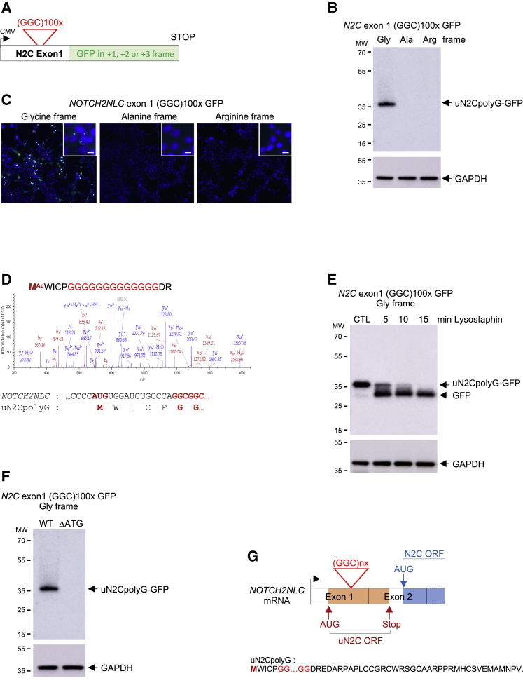
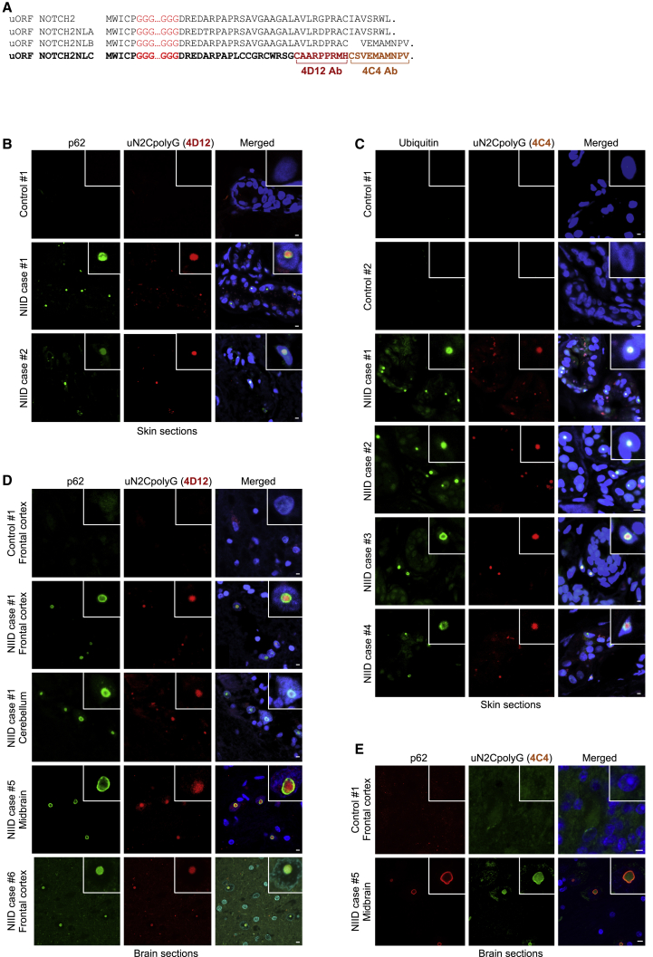
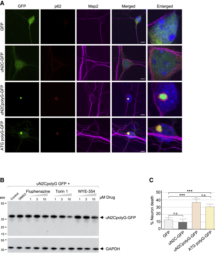
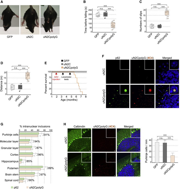
Neuronal intranuclear inclusion disease (NIID) is a neurodegenerative disease characterized by the presence of intranuclear inclusions of unknown origin. NIID is caused by an expansion of GGC repeats in the 5' UTR of the NOTCH2NLC (N2C) gene. We found that these repeats are embedded in a small upstream open reading frame (uORF) (uN2C), resulting in their translation into a polyglycine-containing protein, uN2CpolyG. This protein accumulates in intranuclear inclusions in cell and mouse models and in tissue samples of individuals with NIID. Furthermore, expression of uN2CpolyG in mice leads to locomotor alterations, neuronal cell loss, and premature death of the animals. These results suggest that translation of expanded GGC repeats into a novel and pathogenic polyglycine-containing protein underlies the presence of intranuclear inclusions and neurodegeneration in NIID.

Keywords: RAN translation; genetic diseases; neurodegeneration; polyG; polyglycine; trinucleotide repeat disorder.


Conflict of interest statement

Declaration of interests The authors declare no competing interests.

Figures

None
Graphical abstract
Figure 1
Figure 1
NOTCH2NLC GGC repeats are translated in a polyG-containing protein (A) Schematic of NOTCH2NLC exon 1 with GGC repeats fused to GFP in the glycine, alanine, or arginine frame. (B and C) Immunoblot against GFP (B) or direct fluorescence (C) of HEK293 cells transfected for 24 h with GGC repeats embedded in NOTCH2NLC exon 1 and fused to GFP in the three possible frames. (D) Top panel: N-terminal sequence and corresponding LC-MS/MS spectra of GFP-immunoprecipitated and trypsin-digested protein expressed from uN2C-GFP-transfected HEK293 cells. Bottom panel: nucleotide and amino acid sequences corresponding to the NOTCH2NLC upstream ORF (uN2C) N terminus. (E) Immunoblot against GFP or the glyceraldehyde 3-phosphate dehydrogenase (GAPDH) of lysostaphin-digested proteins extracted from uN2CpolyG-GFP-transfected HEK293 cells. (F) Immunoblot against GFP or the GAPDH of proteins extracted from wild-type or mutant (ΔATG) uN2CpolyG-GFP-transfected HEK293 cells. (G) Top panel: schematic of NOTCH2NLC exons 1 and 2. Bottom panel: amino acid sequence of the NOTCH2NLC upstream ORF (uN2C). See also Figure S1.
Figure 2
Figure 2
The uN2polyG protein is present in NIID intranuclear inclusions (A) Alignment of NOTCH2 and NOTCH2NLA, NOTCH2NLB, and NOTCH2NLC putative upstream ORFs. Brackets indicate the amino acid sequences against which the 4D12 and 4C4 antibodies are directed. (B–E) Immunofluorescence against uN2CpolyG using 4D12 (B and D) or 4C4 (C and E) antibody and p62 (B, D, and E) or ubiquitin (C) on skin (B and C) and brain (D and E) sections of carriers of the NOTCH2NLC-GGC repeat expansion (N2C NIID) or age-matched control individuals. Scale bars, 10 μm. Nuclei were counterstained with DAPI. See also Figure S2 and Table S1.
Figure 3
Figure 3
Expression of uN2CpolyG is toxic in cell culture (A) GFP fluorescence and immunofluorescence against p62 and Map2 of cortical neuronal cell cultures from mouse embryos transduced for 24 h with GFP, uN2C-GFP (12 GGC), uN2CpolyG-GFP (100 GGC), or ATG polyG-GFP (70 GGC) AAV2/PHP.eB. Scale bars, 10 μm. Nuclei were counterstained with DAPI. (B) Immunoblot against GFP or the GAPDH of proteins extracted from uN2CpolyG-GFP-transfected HEK293 cells treated for 15 h with the indicated drugs. (C) Cell viability of cortical neuronal cell cultures from mouse embryos transduced with GFP, uN2C-GFP, uN2CpolyG-GFP, or ATG polyG-GFP AAV2/PHP.eB. Error bars indicate SEM. Student’s t test, ∗∗∗p < 0.001. See also Figures S3, S4, and Table S2.
Figure 4
Figure 4
Expression of uN2CpolyG is pathogenic in animals (A) 60-s tail suspension test shows hind limb clasping in uN2CpolyG-GFP-expressing mice compared with GFP or uN2C-GFP control animals. (B–D) Time before falling from a rotating rod (B), numbers of paw slips and errors in the notched bar test (C), and maximal distance traveled during 30 min in an open field (D) for AAV2/PHP.eB GFP-injected (n = 6), uN2C-GFP-injected (n = 6), and uN2CpolyG-GFP-injected (n = 11) male mice tested 3 months after injection. (E) Kaplan-Meier survival curve of AAV2/PHP.eB GFP-injected (n = 6), uN2C-GFP-injected (n = 6), and uN2CpolyG-GFP-injected (n = 8) male mice. Dates of AAV injection and locomotor tests are indicated by arrows. (F) Immunofluorescence against uN2CpolyG and p62 on cerebellum areas of uN2C-GFP- and uN2CpolyG-GFP-expressing mice sacrificed 2 months after AAV injection. Scale bars, 10 μm. (G) Quantification of p62- or uN2CpolyG-positive intranuclear inclusions in different brain regions of uN2CpolyG-GFP-expressing mice. Brackets indicate the percentage of co-localization between p62- and uN2polyG-positive intranuclear inclusions. N = 3 mice; at least 200 nuclei were counted per brain region and per animal. (H) Left panel: immunofluorescence against uN2CpolyG and calbindin on the cerebellum of uN2C-GFP- and uN2CpolyG-GFP-expressing mice sacrificed 4 months after AAV injection. Scale bars, 20 μm. Right panel: quantification of Purkinje cell numbers in GFP-expressing (n = 4), uN2C-GFP-expressing (n = 4), or uN2CpolyG-GFP-expressing (n = 4) mice. In box-and-whisker plots, box upper and lower limits represent the 25th and 75th percentiles, whiskers represent minimum and maximum values, and a horizontal line across the box represents the median. Bar graphs indicate standard error of the mean (SEM). Student’s t test, ∗∗∗p < 0.001. Nuclei were counterstained with DAPI. See also Figures S5 and S6 and Videos S1, S2, and S3.

Comment in

References

    1. Aspden J.L., Eyre-Walker Y.C., Phillips R.J., Amin U., Mumtaz M.A., Brocard M., Couso J.P. Extensive translation of small Open Reading Frames revealed by Poly-Ribo-Seq. Elife. 2014;3:e03528. - PMC - PubMed
    1. Chen H., Lu L., Wang B., Cui G., Wang X., Wang Y., Raza H.K., Min Y., Li K., Cui Y. Re-defining the clinicopathological spectrum of neuronal intranuclear inclusion disease. Ann. Clin. Transl. Neurol. 2020;7:1930–1941. - PMC - PubMed
    1. Chen Z., Yan Yau W., Jaunmuktane Z., Tucci A., Sivakumar P., Gagliano Taliun S.A., Turner C., Efthymiou S., Ibáñez K., Sullivan R., Genomics England Research Consortium Hardy J, Ryten M, Vandrovcova J, Houlden H. Neuronal intranuclear inclusion disease is genetically heterogeneous. Ann. Clin. Transl. Neurol. 2020;7:1716–1725. - PMC - PubMed
    1. Cortese A., Simone R., Sullivan R., Vandrovcova J., Tariq H., Yau W.Y., Humphrey J., Jaunmuktane Z., Sivakumar P., Polke J. Biallelic expansion of an intronic repeat in RFC1 is a common cause of late-onset ataxia. Nat. Genet. 2019;51:649–658. - PMC - PubMed
    1. Cupidi C., Dijkstra A.A., Melhem S., Vernooij M.W., Severijnen L.A., Hukema R.K., Rozemuller A.J.M., Neumann M., van Swieten J.C., Seelaar H. Refining the Spectrum of Neuronal Intranuclear Inclusion Disease: A Case Report. J. Neuropathol. Exp. Neurol. 2019;78:665–670. - PubMed

Publication types

MeSH terms

Supplementary concepts