. 2021 Apr 23;372(6540):eaba2374.
doi: 10.1126/science.aba2374.

Preventing Engrailed-1 activation in fibroblasts yields wound regeneration without scarring

Affiliations

Preventing Engrailed-1 activation in fibroblasts yields wound regeneration without scarring

Shamik Mascharak et al. Science. .

Abstract

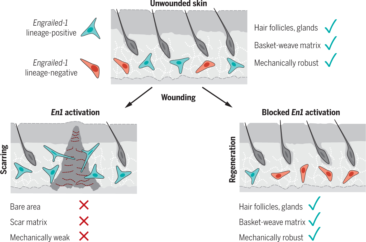
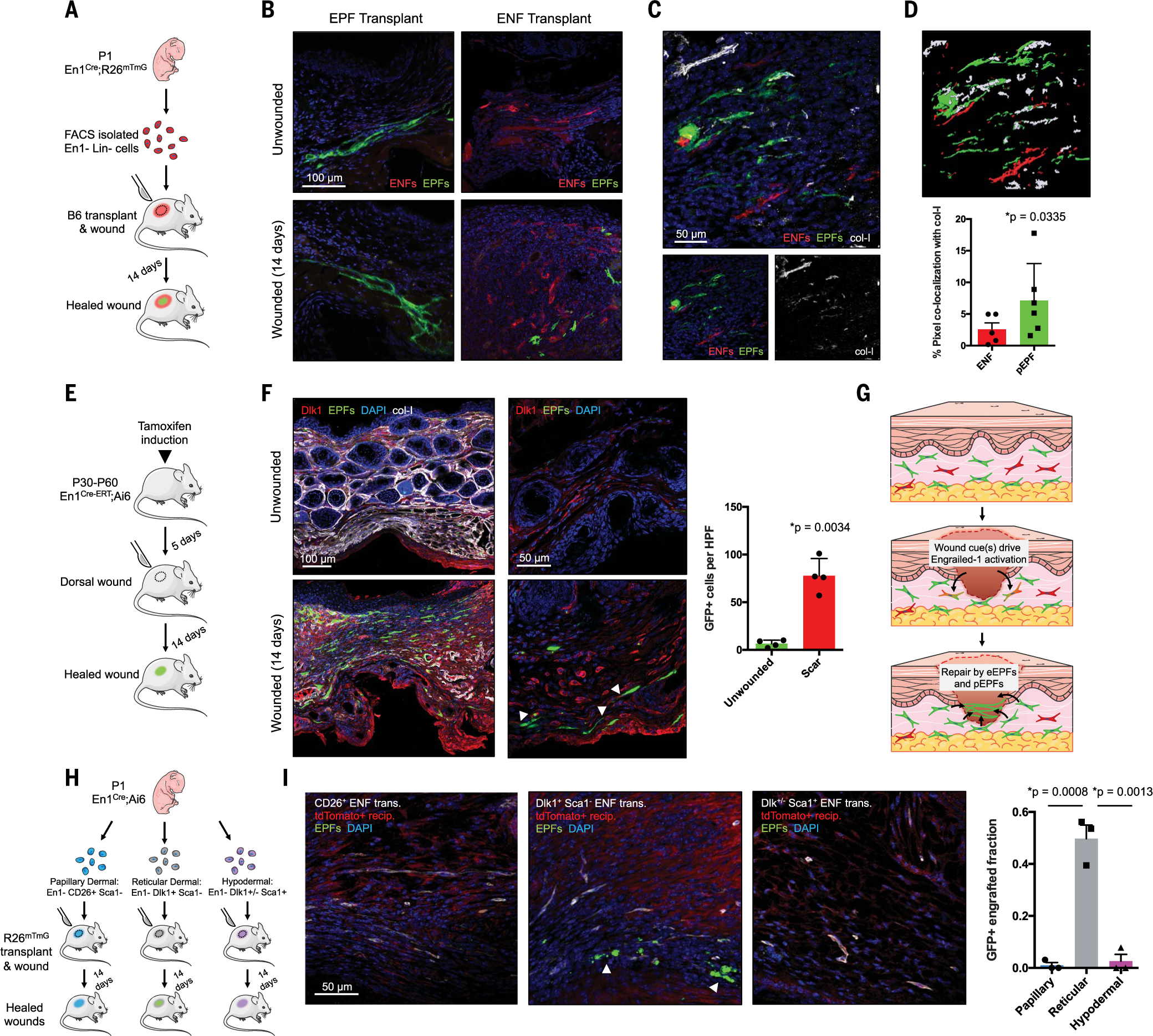
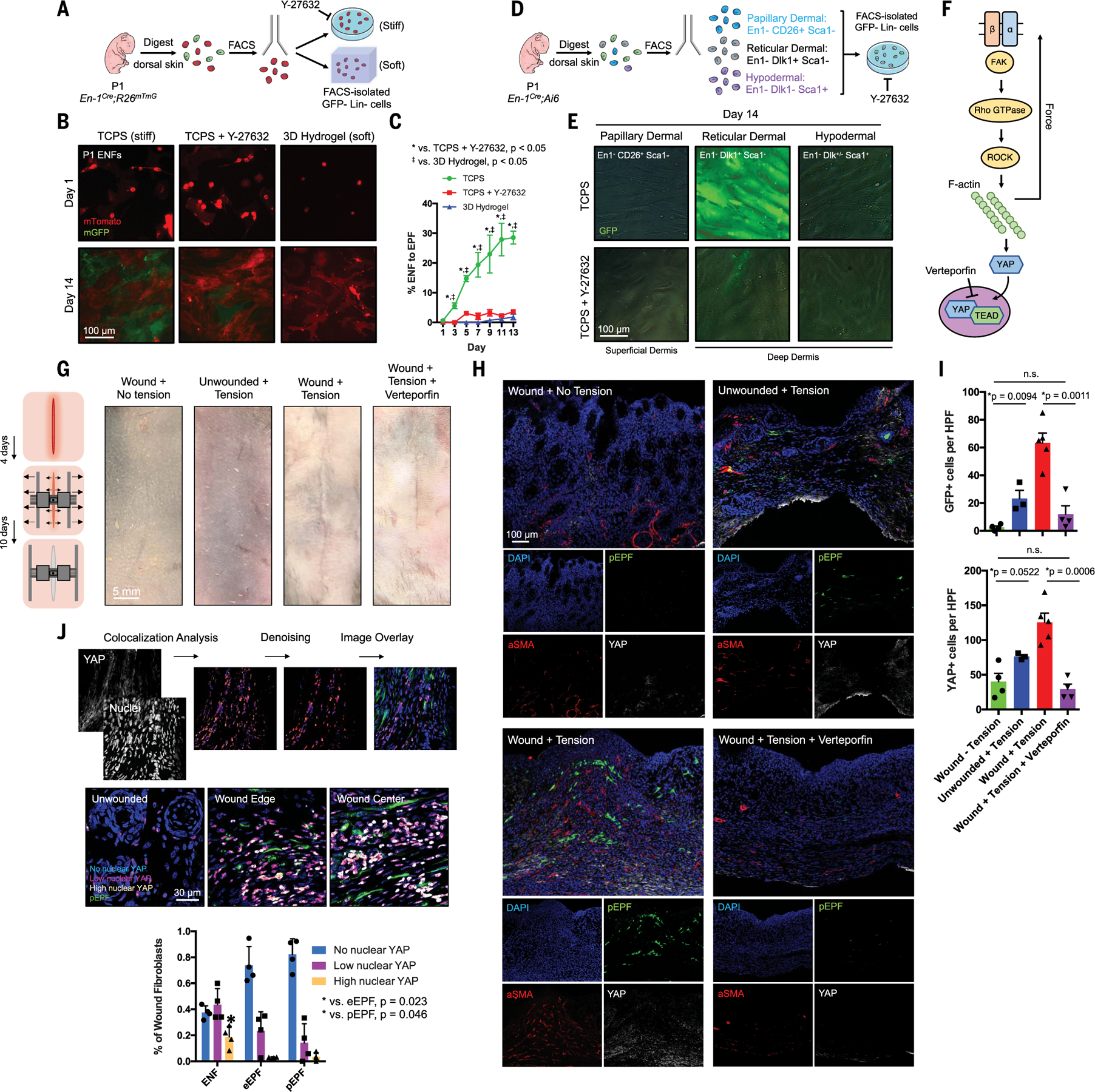
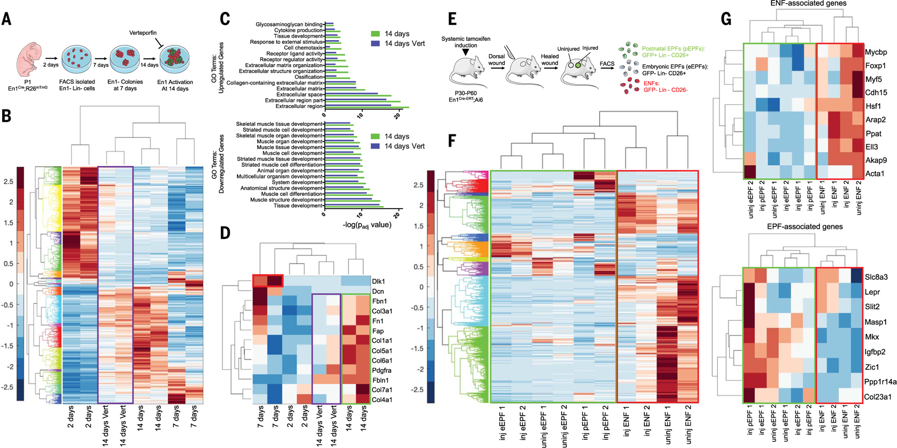
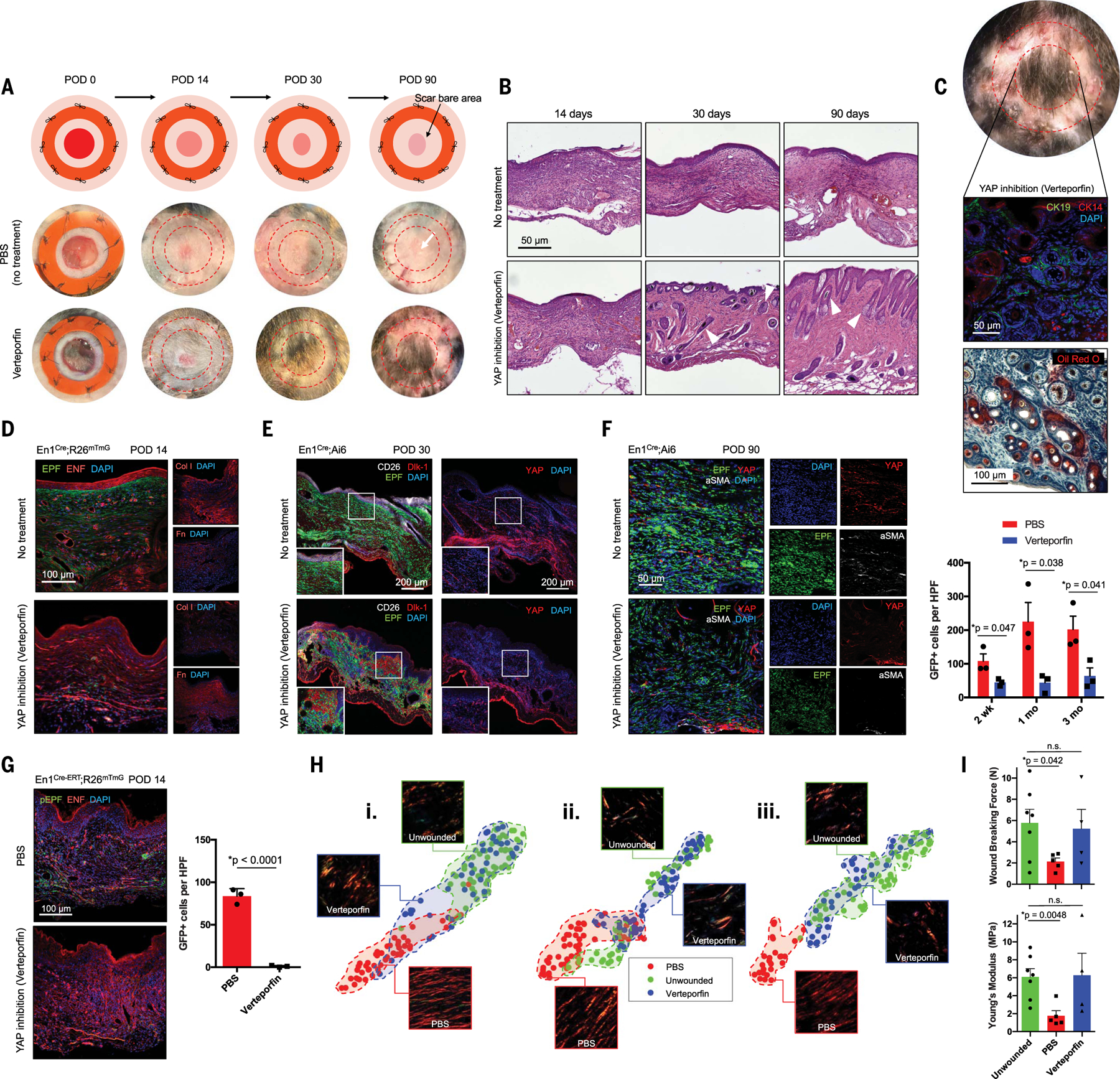
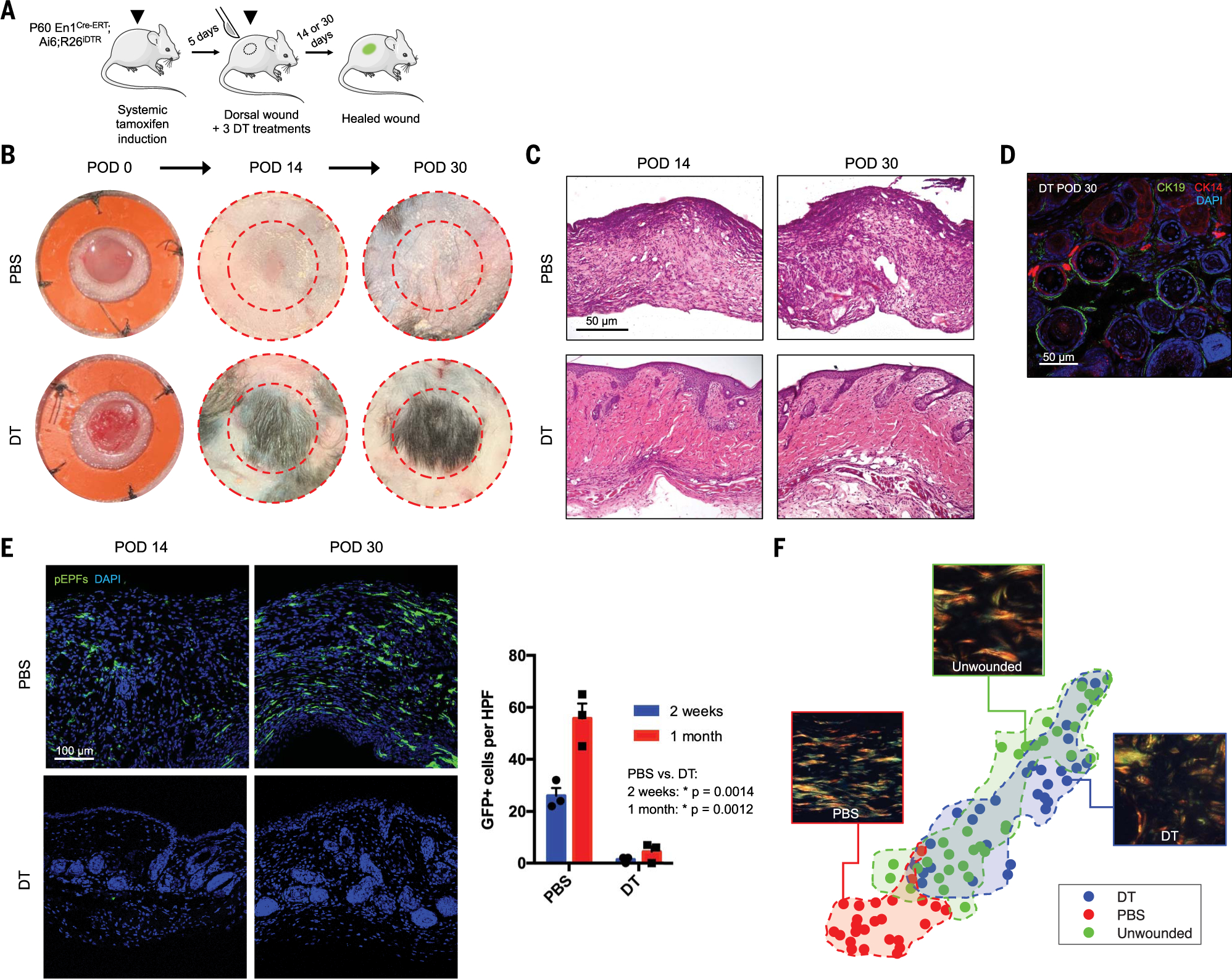
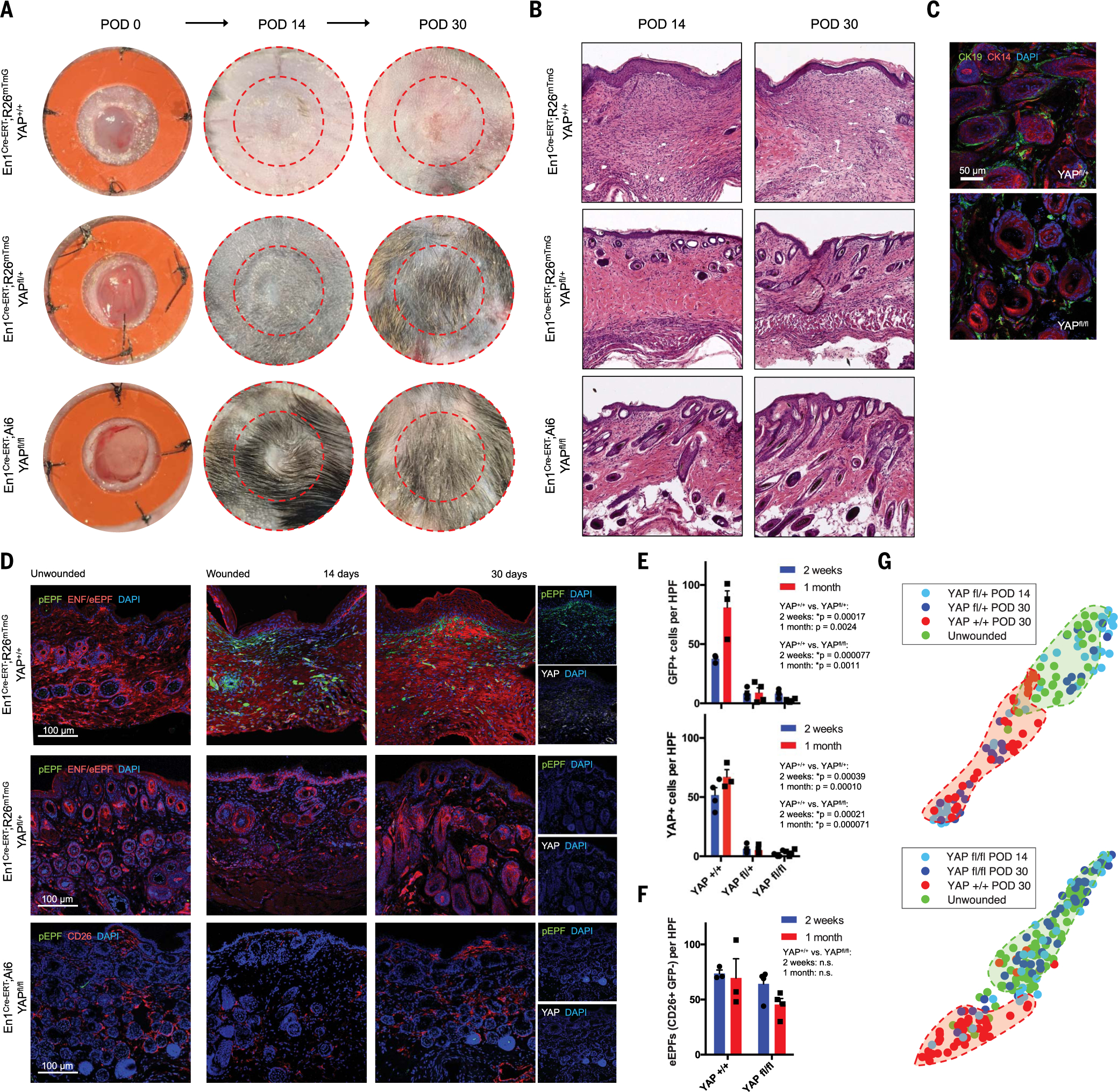
Skin scarring, the end result of adult wound healing, is detrimental to tissue form and function. Engrailed-1 lineage-positive fibroblasts (EPFs) are known to function in scarring, but Engrailed-1 lineage-negative fibroblasts (ENFs) remain poorly characterized. Using cell transplantation and transgenic mouse models, we identified a dermal ENF subpopulation that gives rise to postnatally derived EPFs by activating Engrailed-1 expression during adult wound healing. By studying ENF responses to substrate mechanics, we found that mechanical tension drives Engrailed-1 activation via canonical mechanotransduction signaling. Finally, we showed that blocking mechanotransduction signaling with either verteporfin, an inhibitor of Yes-associated protein (YAP), or fibroblast-specific transgenic YAP knockout prevents Engrailed-1 activation and promotes wound regeneration by ENFs, with recovery of skin appendages, ultrastructure, and mechanical strength. This finding suggests that there are two possible outcomes to postnatal wound healing: a fibrotic response (EPF-mediated) and a regenerative response (ENF-mediated).


Conflict of interest statement

Competing interests: The authors declare no competing interests.

Figures

None
Engrailed-1 activation in skin fibroblasts drives scarring.
After injury, a subset of dermal fibroblasts activates Engrailed-1 (En1) to contribute to scarring (left). Inhibiting postnatal En1 activation, either directly (by ablating En1-activating cells) or indirectly (by blocking mechanical signaling with verteporfin), promotes skin regeneration by En1 lineage-negative fibroblasts, with full recovery of normal hair follicles, glands, matrix ultrastructure, and mechanical strength. Green cells, En1 lineage-positive fibroblasts; red cells, En1 lineage-negative fibroblasts.
Fig. 1.
Fig. 1.. Deep dermal ENFs activate Engrailed-1 and contribute to postnatal scar collagen deposition.
(A) Schematic depicting cell transplantation, engraftment, and wounding experiments. (B) Confocal imaging of transplanted En1-positive fibroblasts (EPFs) and En1-negative fibroblasts (ENFs) before and after wounding [includes 4′,6-diamidino-2-phenylindole (DAPI, blue)]. (C) ENF transplantation and wounding, with postnatal EPFs (pEPFs, green) derived from ENF-to-EPF conversion; immunostaining for type I collagen (col-I). (D) Top: 3D reconstruction of (C). Bottom: Colocalization between col-I and tdTomato (ENF) or GFP (pEPF) signal. (E) Schematic depicting induction and wounding of En1Cre-ERT;Ai6 mice for temporally defined assessment of En1 activation. (F) Left: Skin and wounds of tamoxifen-induced En1Cre-ERT;Ai6 mice; EPFs (white arrowheads) necessarily arose from En1 expression during healing. Immunostaining for Dlk1, col-I. Right: Quantification of GFP+ cells (pEPFs) in unwounded skin or scars; paired two-tailed t test. (G) Proposed mechanism for postnatal En1 activation. Dermal ENFs (red) exposed to wound-specific cues convert to pEPFs, which, with embryonically derived EPFs (eEPFs), mediate scarring. (H) Schematic depicting ENF subtype isolation, transplantation, and wounding. (I) Left: Papillary (CD26+, left), reticular (Dlk1+Sca1, center), and hypodermal (Dlk1+/−Sca1+, right) ENFs in wounded tdTomato+ recipient (red); only reticular ENFs become pEPFs (white arrowheads). Right: Quantification of pEPFs in subtype-engrafted wounds.
Fig. 2.
Fig. 2.. Reticular dermal ENFs activate Engrailed-1 via canonical mechanotransduction signaling in response to in vitro and in vivo substrate mechanics.
(A) Schematic depicting ENF culture on mechanically varied substrates. (B) ENFs (red) cultured on stiff TCPS with or without ROCK inhibitor (Y-27632) or soft hydrogel variably convert to pEPFs (green). (C) Quantification of ENF-to-EPF conversion by condition. (D) Schematic depicting ENF subpopulation culture on TCPS with or without Y-27632. (E) Cultured ENF subtypes show En1 activation (green) only in reticular dermal ENFs on TCPS. (F) Schematic of canonical mechanotransduction signaling. Verteporfin inhibits YAP, the pathway’s transcriptional effector. (G) Left: Schematic depicting wound tension/distraction. Right: Photographs of skin and wounds with or without tension and verteporfin treatment. (H) Fluorescent histology of the four conditions in (G) in En1Cre-ERT;Ai6 showing increased pEPFs (green) with increased tension. Immunofluorescent staining for α-SMA and YAP. (I) Quantification of pEPFs (top) and YAP+ cells (bottom) per 20× high-power field (HPF); n.s., not significant. (J) Top: Pipeline to quantify nuclear YAP. Middle: Overlaid images showing nuclear YAP localization in skin/wound regions. Bottom: Quantification of nuclear YAP levels in ENFs, eEPFs, and pEPFs showing significantly more high–nuclear YAP ENFs than eEPFs or pEPFs.
Fig. 3.
Fig. 3.. Mechanical activation of Dlk1+ ENFs is associated with a fibrotic transcriptional signature.
(A) Schematic of bulk ENF culture over time, with or without verteporfin. (B) Gene expression heatmap and hierarchical clustering for 920 genes significantly up- or down-regulated (by a factor of >4) at 14 days in culture (versus 2 days). Values are shown for 2, 7, or 14 days in culture and 14 days with verteporfin (Vert; purple box). (C) GO term enrichments for significantly up-regulated (top) and down-regulated (bottom) genes from (B), at 14 days with or without Vert. (D) Heatmap showing relative expression of selected genes previously implicated in fibrosis/ECM deposition. Dlk1 expression was up-regulated in ENFs at 7 days (red box). Profibrotic/matrix genes were up-regulated at 14 days (green box) but mitigated by Vert (purple box). (E) Schematic depicting isolation of skin and scar pEPFs, eEPFs, and ENFs for RNA-seq. (F) Heatmap and hierarchical clustering of 1138 genes significantly up- or down-regulated in ENFs, eEPFs, or pEPFs in wounds (inj) versus skin (uninj). (G) Heatmaps showing relative expression of selected genes previously associated with ENF (top) or EPF (bottom) identity. Green boxes, EPF populations; red boxes, ENFs.
Fig. 4.
Fig. 4.. Mechanotransduction inhibition in vivo results in scarless wound healing via regeneration.
(A) Schematic (top) and gross photographs of dorsal excisional wounds treated with PBS (control) or verteporfin. Surrounding bare area is where splint was attached and removed before harvest (red dashed circles). (B) H&E histology of control- and verteporfin-treated wounds. White arrowheads indicate dermal appendage–like morphology. (C) By POD 90, verteporfin-treated wounds regenerate HF/appendages, grossly (top) and histologically: middle, immunostaining for HF/SG markers CK14/19; bottom, Oil Red O staining (red) for SG. (D to F) Fluorescent histology of control- or verteporfin-treated wounds in indicated mice at POD 14 (D), 30 (E), and 90 (F) with immunostaining for ECM proteins (col-I, Fn) and fibroblast/mechanotransduction markers (CD26, Dlk1, YAP, α-SMA). (F) Right: EPFs per HPF in PBS- and verteporfin-treated wounds over time. (G) Left: Fluorescent histology of control- and verteporfin-treated En1Cre-ERT;R26mTmG wounds. Right: Quantification of pEPFs per HPF. (H) t-distributed stochastic neighbor embedding (t-SNE) plots visualizing ECM ultrastructural properties for skin and PBS- or verteporfin-treated wounds at POD 14 (i), 30 (ii), and 90 (iii); clusters for each condition (PBS-treated, unwounded, or verteporfin-treated) are highlighted by shaded regions. (I) Wound breaking force (top; unwounded versus verteporfin, P = 0.8057) and Young’s modulus (bottom; unwounded versus verteporfin, P = 0.9287) calculated for unwounded skin and PBS- or verteporfin-treated wounds.
Fig. 5.
Fig. 5.. Targeted ablation of Engrailed-1–expressing fibroblasts yields skin wound regeneration.
(A) Schematic of En1Cre-ERT;Ai6;R26iDTR tamoxifen induction, wounding, and diphtheria toxin (DT) treatment to ablate pEPFs generated in response to wounding. (B and C) Gross photographs (B) and H&E histology (C) of wounds treated with PBS (top) or DT (bottom) over time. (D) DT-treated wounds at POD 30 with immunofluorescent staining for CK14/19 (HF/SG). (E) Left: Fluorescent histology of PBS- and DT-treated wounds at POD 14 and 30, showing pEPFs (GFP+). Right: Quantification of pEPFs per 20× HPF. (F) t-SNE plots visualizing 26 ECM ultrastructural properties for unwounded skin and PBS- or DT-treated wounds at POD 30; shaded regions highlight clusters of the three conditions.
Fig. 6.
Fig. 6.. YAP knockout abrogates postnatal Engrailed-1 expression to promote skin wound regeneration.
(A and B) Gross photographs (A) and H&E histology (B) of YAP+/+ (top), YAPfl/+ (middle), and YAPfl/fl (bottom) wounds over time. (C) Immunofluorescent histology for CK14/19 (HF/SG) in POD 30 YAPfl/+ (top) and YAPfl/fl (bottom) wounds. (D) Immunofluorescent histology of skin (left) and YAP+/+ (top), YAPfl/+ (middle), and YAPfl/fl (bottom) wounds at POD 14 and 30. Rightmost panels show pEPFs (GFP+) and YAP immunostaining. (E) Quantification of pEPFs (top) and YAP+ cells (bottom) per 20× HPF in YAP+/+, YAPfl/+, and YAPfl/fl wounds at POD 14 and 30. (F) Quantification of eEPFs (CD26+GFP) in YAP+/+ and YAPfl/fl wounds. (G) t-SNE plots visualizing ECM ultrastructural properties for unwounded skin versus YAPfl/+ (top) and YAPfl/fl (bottom) wounds; shaded regions highlight clusters of the four conditions.

Comment in

  • Healing without scarring.
    Konieczny P, Naik S. Konieczny P, et al. Science. 2021 Apr 23;372(6540):346-347. doi: 10.1126/science.abi5770. Science. 2021. PMID: 33888629 No abstract available.

References

    1. Gurtner GC, Werner S, Barrandon Y, Longaker MT, Wound repair and regeneration. Nature 453, 314–321 (2008). doi:10.1038/nature07039 - DOI - PubMed
    1. Bayat A, McGrouther DA, Ferguson MW, Skin scarring. BMJ 326, 88–92 (2003). doi:10.1136/bmj.326.7380.88 - DOI - PMC - PubMed
    1. Eming SA, Martin P, Tomic-Canic M, Wound repair and regeneration: Mechanisms, signaling, and translation. Sci. Transl. Med. 6, 265sr6 (2014). doi:10.1126/scitranslmed.3009337 - DOI - PMC - PubMed
    1. Sun BK, Siprashvili Z, Khavari PA, Advances in skin grafting and treatment of cutaneous wounds. Science 346, 941–945 (2014). doi:10.1126/science.1253836 - DOI - PubMed
    1. Griffin MF, desJardins-Park HE, Mascharak S, Borrelli MR, Longaker MT, Understanding the impact of fibroblast heterogeneity on skin fibrosis. Dis. Model. Mech 13, dmm044164 (2020). doi:10.1242/dmm.044164 - DOI - PMC - PubMed

Publication types

MeSH terms