. 2021 Apr 22;11(1):8802.
doi: 10.1038/s41598-021-88026-9.

Rapid uropathogen identification using surface enhanced Raman spectroscopy active filters

Affiliations

Rapid uropathogen identification using surface enhanced Raman spectroscopy active filters

Simon D Dryden et al. Sci Rep. .

Abstract

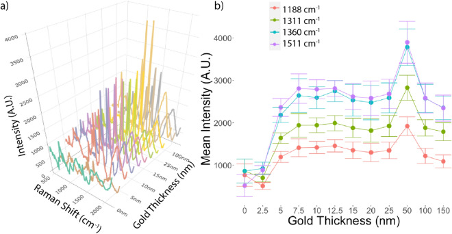
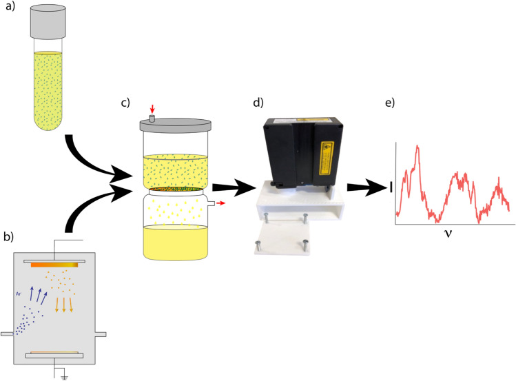
Urinary tract infection is one of the most common bacterial infections leading to increased morbidity, mortality and societal costs. Current diagnostics exacerbate this problem due to an inability to provide timely pathogen identification. Surface enhanced Raman spectroscopy (SERS) has the potential to overcome these issues by providing immediate bacterial classification. To date, achieving accurate classification has required technically complicated processes to capture pathogens, which has precluded the integration of SERS into rapid diagnostics. This work demonstrates that gold-coated membrane filters capture and aggregate bacteria, separating them from urine, while also providing Raman signal enhancement. An optimal gold coating thickness of 50 nm was demonstrated, and the diagnostic performance of the SERS-active filters was assessed using phantom urine infection samples at clinically relevant concentrations (105 CFU/ml). Infected and uninfected (control) samples were identified with an accuracy of 91.1%. Amongst infected samples only, classification of three bacteria (Escherichia coli, Enterococcus faecalis, Klebsiella pneumoniae) was achieved at a rate of 91.6%.


Conflict of interest statement

The authors declare no competing interests.

Figures

Figure 1
Figure 1
Effect of gold coating thickness on Rhodamine 6G Raman spectra. (a) Mean Raman Spectra of Rhodamine 6G on PVDF filters with differing gold coating thicknesses. (b) Mean intensity with standard deviation (error bar) of the four most prominent peaks in the Rhodamine 6G spectra (1188, 1311, 1360 and 1511 cm−1) for differing gold coating thicknesses. Thickness greater than 2.5 nm provided significant enhancement of all 4 prominent peaks, with the greatest enhancement seen with filters with a 50 nm gold coating.
Figure 2
Figure 2
(a) Mean normalized Raman spectra and standard deviations (shaded areas) for infected (red) and uninfected (blue) urine samples captured on SERS-active filters. (b) Mean spectrum with standard deviation (ribbon) of infected samples centered and scaled on uninfected controls demonstrating discriminatory peaks at 710, 855, 1004, 1235, 1375 and 1505 cm-1.
Figure 3
Figure 3
Identification of bacteria based on Raman spectra captured on SERS-active filters. (a) Mean Raman spectra with standard deviations (shaded areas) for Escherichia coli (red), Enterococcus faecalis (green) and Klebsiella pneumoniae (blue) captured on SERS-active filters. b) Spectra of individual species scaled and centered on one another demonstrating discriminatory peaks at 710, 855, 1004, 1235 and 1375 cm-1 (indicated by dotted lines).
Figure 4
Figure 4
PC-LDA performed on Raman spectra from urine samples inoculated with Escherichia coli (red circle), Enterococcus faecalis (green triangle) and Klebsiella pneumoniae (blue square).
Figure 5
Figure 5
Methods overview. (a) Phantom urine samples were prepared by inoculating uropathogens (illustrated as blue ovals) into urine samples from healthy volunteers. (b) SERS-active filters were prepared by applying a 50 nm gold coating to polyvinylidene fluoride membrane filters using physical vapour deposition. (c) Vacuum filtration captured the uropathogens from urine and applied them directly to the SERS-enhancing surface (Red arrows indicate direction of vacuum). (d) Raman spectra were collected with a handheld spectrometer supported by a custom 3D-printed holder. (e) The spectra were plotted and analyzed (I—intensity, ν—wavenumber).

References

    1. Ozturk R, Murt A. Epidemiology of urological infections: A global burden. World J. Urol. 2020;38:2669–2679. doi: 10.1007/s00345-019-03071-4. - DOI - PubMed
    1. Stamm WE, Norrby SR. Urinary tract infections: Disease panorama and challenges. J. Infect. Dis. 2001;183:S1–S4. doi: 10.1086/318850. - DOI - PubMed
    1. Flores-Mireles AL, Walker JN, Caparon M, Hultgren SJ. Urinary tract infections: Epidemiology, mechanisms of infection and treatment options. Nat. Rev. Microbiol. 2015;13:269–284. doi: 10.1038/nrmicro3432. - DOI - PMC - PubMed
    1. Foxman B. Urinary tract infection syndromes. Infect. Dis. Clin. North Am. 2014;28:1–13. doi: 10.1016/j.idc.2013.09.003. - DOI - PubMed
    1. Scholes D, et al. Risk factors associated with acute pyelonephritis in healthy women. Ann. Intern. Med. 2005;142:20–27. doi: 10.7326/0003-4819-142-1-200501040-00008. - DOI - PMC - PubMed

Publication types

MeSH terms