. 2021 Apr 22;11(1):8744.
doi: 10.1038/s41598-021-88157-z.

C subunit of the ATP synthase is an amyloidogenic calcium dependent channel-forming peptide with possible implications in mitochondrial permeability transition

Affiliations

C subunit of the ATP synthase is an amyloidogenic calcium dependent channel-forming peptide with possible implications in mitochondrial permeability transition

Giuseppe Federico Amodeo et al. Sci Rep. .

Abstract

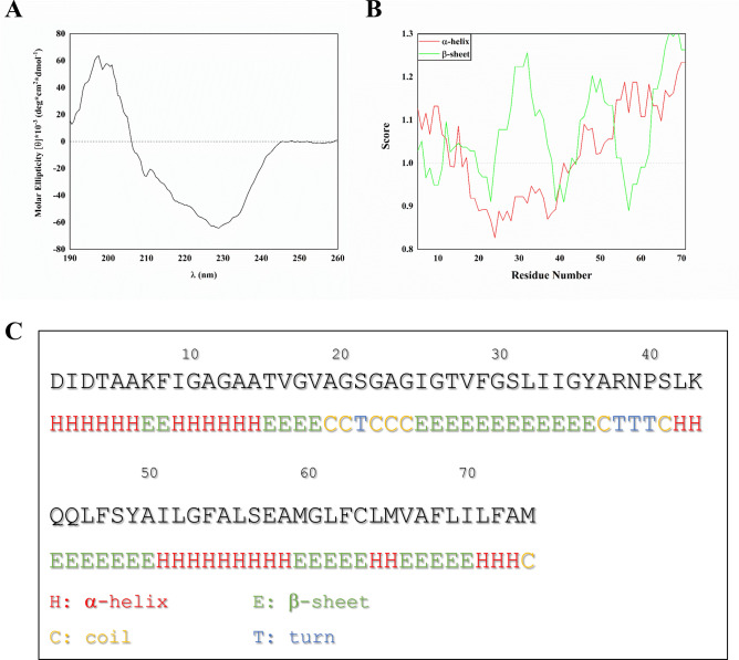
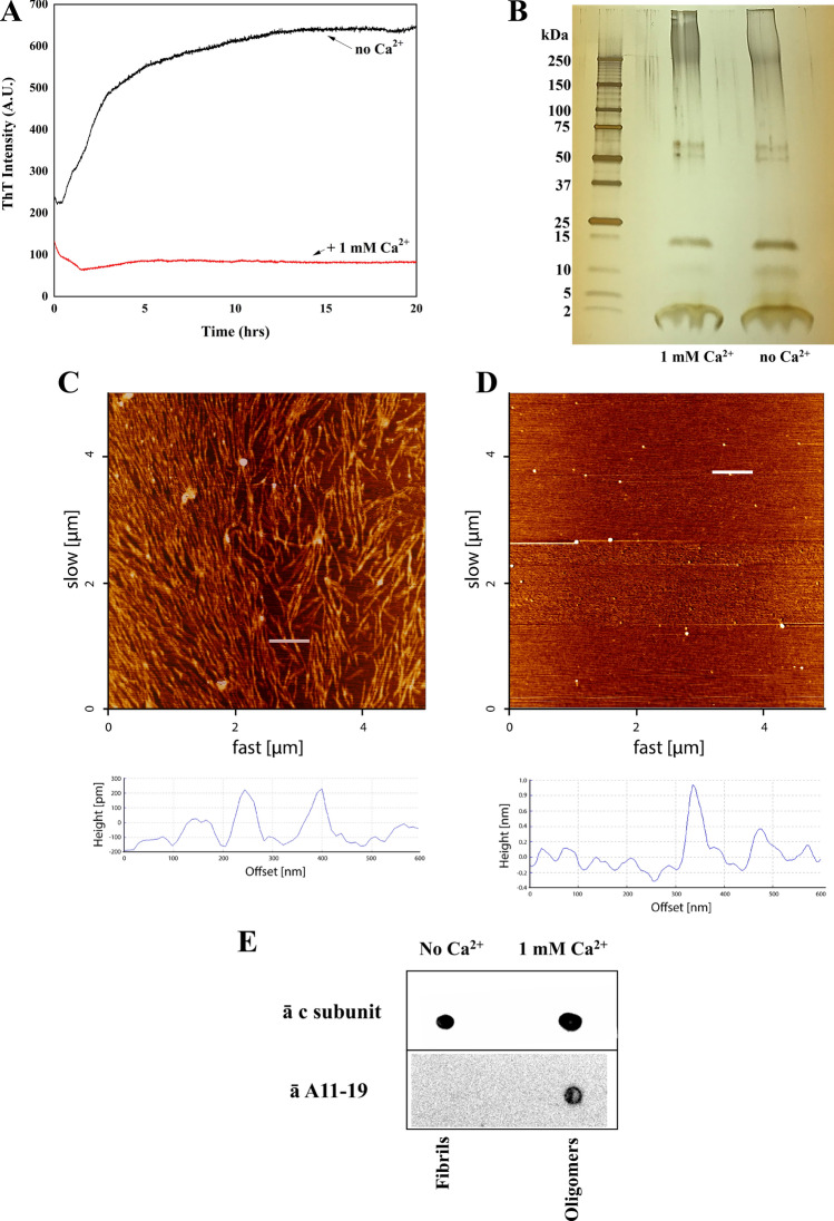
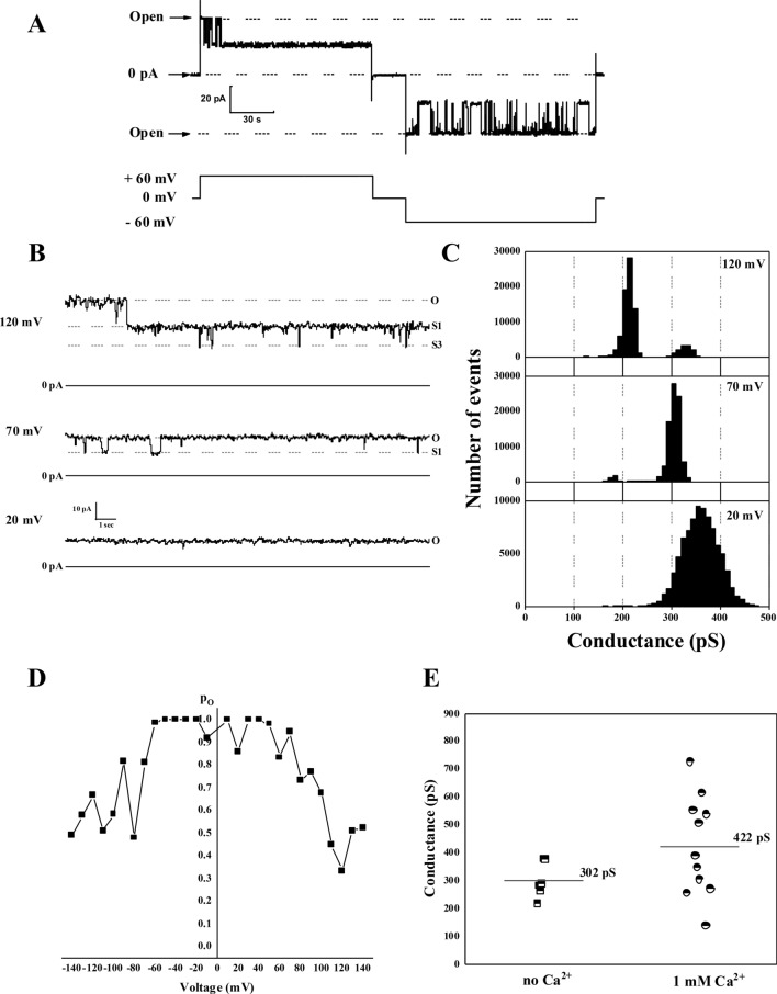
The c subunit is an inner mitochondrial membrane (IMM) protein encoded by three nuclear genes. Best known as an integral part of the F0 complex of the ATP synthase, the c subunit is also present in other cytoplasmic compartments in ceroid lipofuscinoses. Under physiological conditions, this 75 residue-long peptide folds into an α-helical hairpin and forms oligomers spanning the lipid bilayer. In addition to its physiological role, the c subunit has been proposed as a key participant in stress-induced IMM permeabilization by the mechanism of calcium-induced permeability transition. However, the molecular mechanism of the c subunit participation in IMM permeabilization is not completely understood. Here we used fluorescence spectroscopy, atomic force microscopy and black lipid membrane methods to gain insights into the structural and functional properties of unmodified c subunit protein that might make it relevant to mitochondrial toxicity. We discovered that c subunit is an amyloidogenic peptide that can spontaneously fold into β-sheets and self-assemble into fibrils and oligomers in a Ca2+-dependent manner. C subunit oligomers exhibited ion channel activity in lipid membranes. We propose that the toxic effects of c subunit might be linked to its amyloidogenic properties and are driven by mechanisms similar to those of neurodegenerative polypeptides such as Aβ and α-synuclein.


Conflict of interest statement

The authors declare no competing interests.

Figures

Figure 1
Figure 1
C subunit forms β-sheet structures. (A) CD spectrum of the 50 µM c subunit in 2% Genapol and PBS; (B) Bioinformatic prediction of the probability score of the secondary structure of the c subunit based on its primary sequence according to the Chou and Fasman method; (C) Most probable predicted structure per individual residue (Created with OriginPro 2021, OriginLab Corp. Northampton, MA).
Figure 2
Figure 2
Aggregation properties of the c subunit. (A) Thioflavin T fluorescence spectra showing calcium dependence of c subunit aggregation (created with OriginPro 2021, OriginLab Corp. Northampton, MA); (B) SDS PAGE of the c subunit showing presence of oligomeric and monomeric forms; (C) 5 × 5 µm AFM image of c subunit fibrils after incubation at 37 °C for 4 h in 2% Genapol and PBS; (D) 5 × 5 µm AFM image of the c subunit oligomers (arrows) after incubation at 37 °C for 4 h and deposited on mica. Below each image is provided the height profile of the indicated white line; (E) Dot blot of the c subunit incubated with anti c subunit and A11-19 antibodies with and without Ca2+ showing the ability of the protein to form cross-β oligomers (full-length blots are presented in Supplementary Figure 1).
Figure 3
Figure 3
Ion channel activity of c subunit oligomers. (A) Representative current trace of oligomers showing typical channel behaviour with frequent transitions between fully open and lower conductance states; (B) Representative c subunit channel activity at different voltages; (C) All points histogram corresponding to the trace shown at panel (B); (D) Voltage dependence of the open probability of the c subunit channel; (E) Channel open state conductance values of the c subunit channels from multiple independent experiments alone (n = 5) and in the presence of Ca2+ (n = 11) (Created with OriginPro 2021, OriginLab Corp. Northampton, MA).
Figure 4
Figure 4
Function of c subunit in physiological and pathological mitochondria. The c subunit is encoded by three nuclear genes: ATP5G1, ATP5G2 and ATP5G3. Each of these genes, when translated, express the same mature sequence of the c subunit with differing signal peptides (A). Predictions based on the immature sequences of the c subunit isoforms indicate that the protein may be imported into mitochondria through the TIM/TOM complex (B). In physiological conditions (C), c subunit monomers fold into their native α-helical hairpin conformation and oligomerize into the c-ring of the ATPase. Conversely, in pathological conditions (E), the increased levels of Ca2+ influence the misfolding of the newly imported peptides causing them to oligomerize into a cross-β conformation (F) that contributes to PTP through permeabilizing the IMM (Created with Biorender.com).

References

    1. Walker JE. The ATP synthase: The understood, the uncertain and the unknown. Biochem. Soc. Trans. 2013;41:1–16. doi: 10.1042/BST20110773. - DOI - PubMed
    1. Palmer DN. The relevance of the storage of subunit c of ATP synthase in different forms and models of Batten disease (NCLs) Biochim. Biophys. Acta. 1852;2287–2291:2015. doi: 10.1016/j.bbadis.2015.06.014. - DOI - PubMed
    1. Palmer DN, et al. Mitochondrial ATP synthase subunit c storage in the ceroid-lipofuscinoses (Batten disease) Am. J. Med. Genet. 1992;42:561–567. doi: 10.1002/ajmg.1320420428. - DOI - PubMed
    1. Alavian KN, et al. An uncoupling channel within the c-subunit ring of the F1FO ATP synthase is the mitochondrial permeability transition pore. Proc. Natl. Acad. Sci. U.S.A. 2014;111:10580–10585. doi: 10.1073/pnas.1401591111. - DOI - PMC - PubMed
    1. Bonora M, et al. Role of the c subunit of the FO ATP synthase in mitochondrial permeability transition. Cell Cycle. 2013;12:674–683. doi: 10.4161/cc.23599. - DOI - PMC - PubMed

Publication types

MeSH terms

Substances