

Methanolic Fenugreek Seed Extract Induces p53-Dependent Mitotic Catastrophe in Breast Cancer Cells, Leading to Apoptosis
- PMID: 33889009
- PMCID: PMC8057839
- DOI: 10.2147/JIR.S300025
Methanolic Fenugreek Seed Extract Induces p53-Dependent Mitotic Catastrophe in Breast Cancer Cells, Leading to Apoptosis
Abstract
Purpose: The plant Trigonella foenum-graecum, well-known as fenugreek, has been shown to control type-2 diabetes, the level of cholesterol, inflammation of wounds, disorders related to gastrointestinal tracts, and cancer as well. The present study aimed to evaluate the anti-cancer potential of methanolic fenugreek seed extract (FSE) and its possible molecular mechanism of action in breast cancer cells.
Methods: The anticancer potential of FSE was evaluated in MCF-7 and SK-BR3 breast cancer cells through various cellular assays after selecting the IC10, IC25, IC35, and IC50 doses by the cell cytotoxicity assay. Furthermore, the oral acute toxicity of FSE was examined in mice, according to the guidelines of the Organization for Economic Co-operation and Development (OECD).
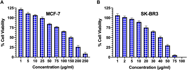
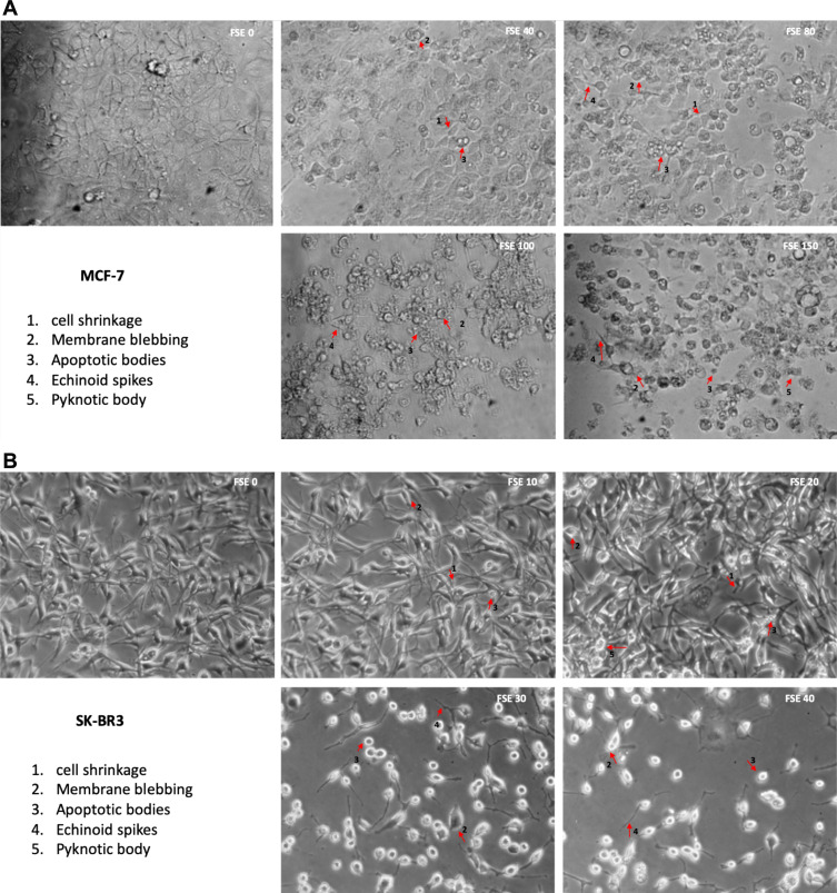
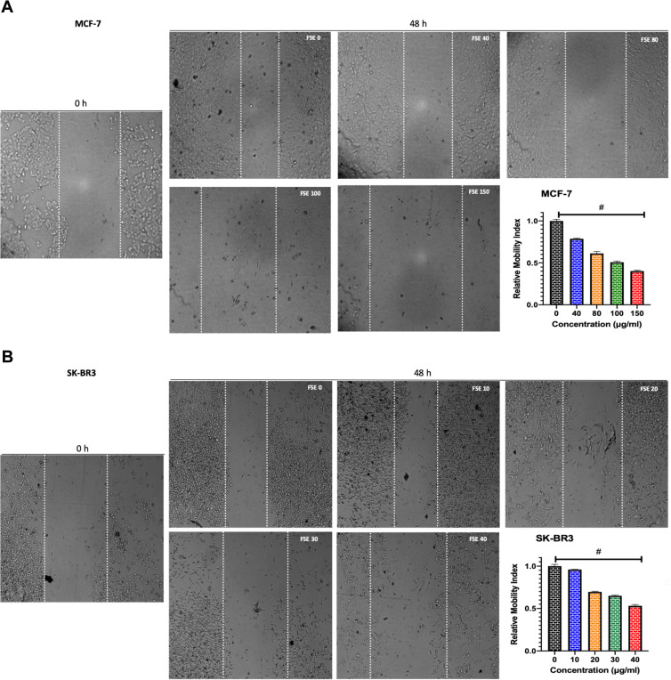
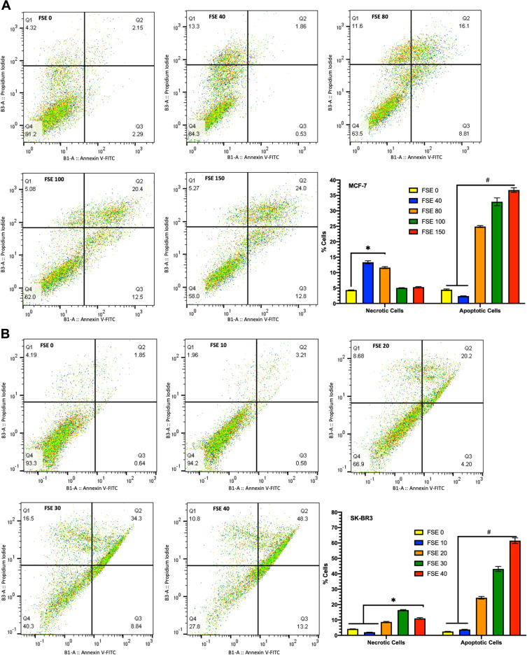
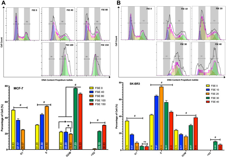
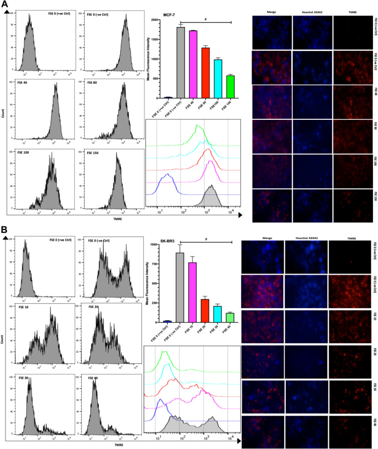
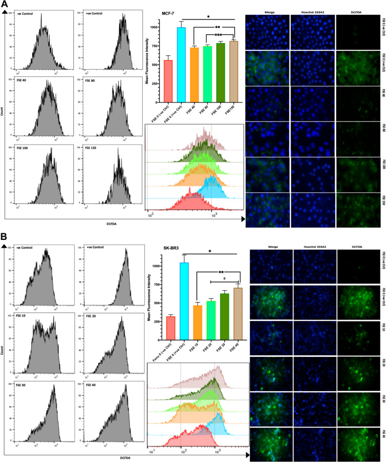
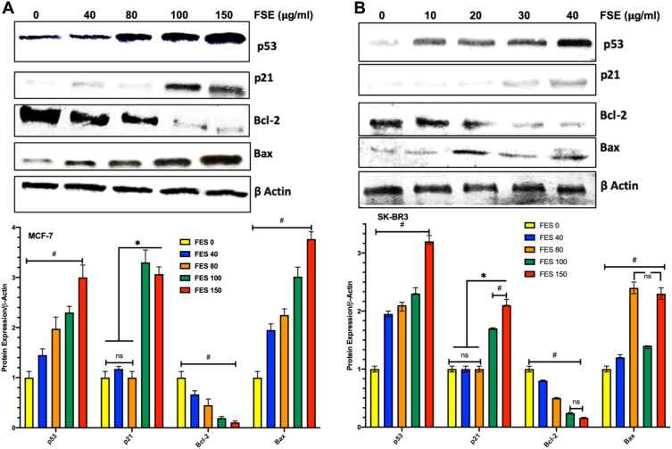
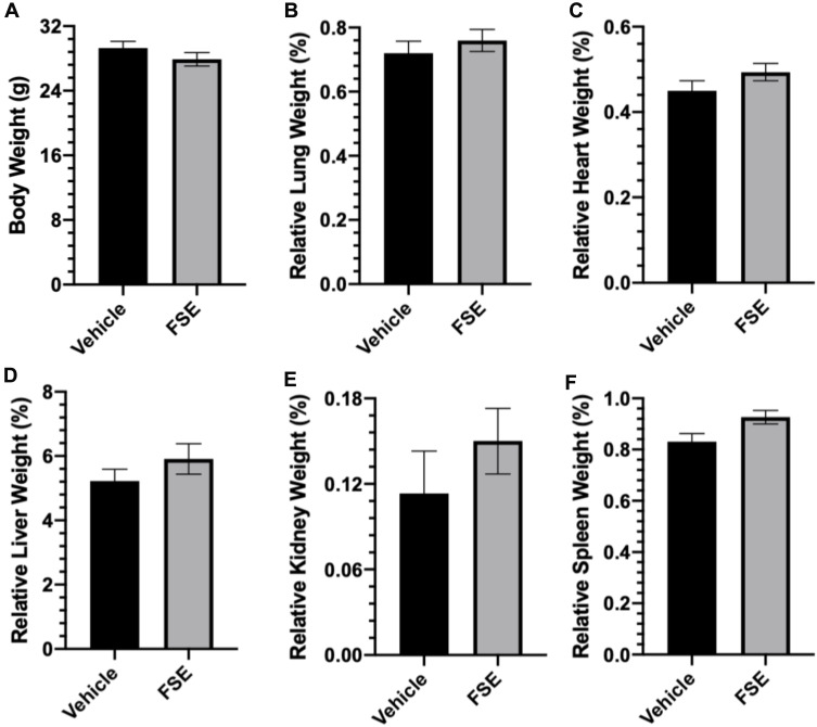
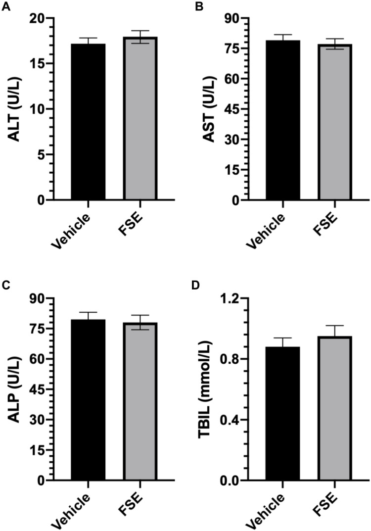
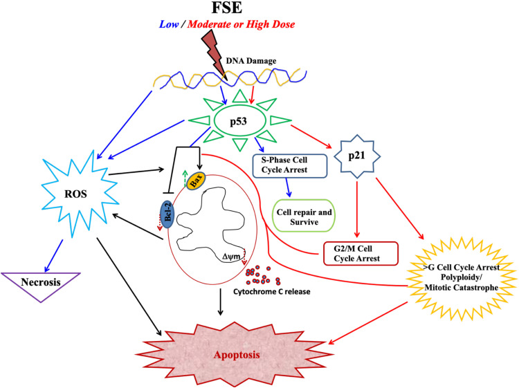
Results: FSE exhibited dose-dependent cytotoxicity, as the IC50 was found to be 150 and 40 μg/mL for MCF-7 and SK-BR3 breast cancer cells, respectively. The cytological observations showed the typical apoptotic morphology in both of the breast cancer cells upon treatment with FSE, as it inhibited the migration and adhesion, in a dose-dependent manner. The flow cytometry analysis revealed that FSE induced a significant shift from G2/M, and polyploidy (>G) at higher concentrations that suggested the activation of p53-mediated mitotic catastrophe, consequently leading to apoptosis. FSE induced a significant increase in the mitochondrial depolarization, ROS as well as a Bax/Bcl-2 ratio, and also exhibited the mitochondrial associated p53 signaling pathway. The in vivo acute toxicity data revealed that the oral administration of FSE did not induce any toxic effect in mice.
Conclusion: This study, for the first time, reports the mechanistic details of the anti-cancer potential of FSE. It requires a detailed analysis to understand the effect of FSE to induce the apoptosis through the multiple signaling pathways at varying concentrations. The nontoxic effect of FSE in mice suggests to utilize it safely for pharmaceutical formulations in different cancer systems.
Keywords: apoptosis; breast cancer cells; fenugreek seed extract; mitochondria-associated pathway; mitotic catastrophe; oral acute toxicity; p53 signaling.
© 2021 Alrumaihi et al.
Conflict of interest statement
The authors report no conflicts of interest in this work.
Figures

Similar articles
-
Hepatoprotective effect of Trigonella foenum graecum against ethanol-induced cell death in human liver cells (HepG2 and Huh7).Mol Biol Rep. 2022 Apr;49(4):2765-2776. doi: 10.1007/s11033-021-07088-0. Epub 2022 Jan 22. Mol Biol Rep. 2022. PMID: 35064405
-
Anticancer activity of Trigonella Foenumgraecum (fenugreek) seed extract by inducing apoptosis in pancreatic cancer cell.Am J Transl Res. 2025 Feb 15;17(2):832-843. doi: 10.62347/PGLT6191. eCollection 2025. Am J Transl Res. 2025. PMID: 40092083 Free PMC article.
-
Preclinical and Molecular Docking Insights into the Chemopreventive Role of Fenugreek Seed Extract in a Murine Model of Colorectal Cancer.Pharmaceuticals (Basel). 2025 Mar 28;18(4):490. doi: 10.3390/ph18040490. Pharmaceuticals (Basel). 2025. PMID: 40283928 Free PMC article.
-
Preclinical Toxicological Evaluation of IDM01: The Botanical Composition of 4-Hydroxyisoleucine- and Trigonelline-based Standardized Fenugreek Seed Extract.Pharmacognosy Res. 2017 Apr-Jun;9(2):138-150. doi: 10.4103/0974-8490.204649. Pharmacognosy Res. 2017. PMID: 28539737 Free PMC article.
-
Anticancer potential of Trigonella foenum graecum: Cellular and molecular targets.Biomed Pharmacother. 2017 Jun;90:479-491. doi: 10.1016/j.biopha.2017.03.071. Epub 2017 Apr 6. Biomed Pharmacother. 2017. PMID: 28391170 Review.
Cited by
-
Fenugreek inhibits cathepsin G activity and suppresses the progression of malignant phenotypes in MCF-7 cells.Biochem Biophys Rep. 2025 Apr 21;42:102021. doi: 10.1016/j.bbrep.2025.102021. eCollection 2025 Jun. Biochem Biophys Rep. 2025. PMID: 40524916 Free PMC article.
-
Spice-Derived Phenolic Compounds: Potential for Skin Cancer Prevention and Therapy.Molecules. 2023 Aug 25;28(17):6251. doi: 10.3390/molecules28176251. Molecules. 2023. PMID: 37687080 Free PMC article. Review.
-
Apoptotic effect of thymoquinone on OVCAR3 cells via the P53 and CASP3 activation.Acta Cir Bras. 2024 Nov 8;39:e399224. doi: 10.1590/acb399224. eCollection 2024. Acta Cir Bras. 2024. PMID: 39536185 Free PMC article.
-
Revisiting Trigonella foenum-graecum L.: Pharmacology and Therapeutic Potentialities.Plants (Basel). 2022 May 29;11(11):1450. doi: 10.3390/plants11111450. Plants (Basel). 2022. PMID: 35684222 Free PMC article. Review.
-
Spices and culinary herbs for the prevention and treatment of breast cancer: A comprehensive review with mechanistic insights.Cancer Pathog Ther. 2024 Jul 6;3(3):197-214. doi: 10.1016/j.cpt.2024.07.003. eCollection 2025 May. Cancer Pathog Ther. 2024. PMID: 40458304 Free PMC article. Review.
References
-
- Kaefer CM, Milner JA. Herbs and spices in cancer prevention and treatment. In: Benzie IFF, Wachtel-Galor S, editors. Herbal Medicine: Biomolecular and Clinical Aspects. 2nd edition. Boca Raton (FL): CRC Press/Taylor & Francis; 2011:Chapter 17. Available from: www.ncbi.nlm.nih.gov/books/NBK92774/.
-
- Birt DF, Pelling JC, Nair S, et al. Diet intervention for modifying cancer risk. Prog Clin Biol Res. 1996;395:223–234. - PubMed
LinkOut - more resources
Full Text Sources
Other Literature Sources
Research Materials
Miscellaneous
