

Transneuronal Degeneration in the Brain During Glaucoma
- PMID: 33889083
- PMCID: PMC8055862
- DOI: 10.3389/fnagi.2021.643685
Transneuronal Degeneration in the Brain During Glaucoma
Abstract
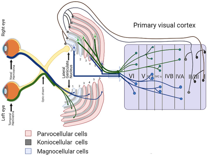
The death of retinal ganglion cells (RGCs) is a key factor in the pathophysiology of all types of glaucoma, but the mechanism of pathogenesis of glaucoma remains unclear. RGCs are a group of central nervous system (CNS) neurons whose soma are in the inner retina. The axons of RGCs form the optic nerve and converge at the optic chiasma; from there, they project to the visual cortex via the lateral geniculate nucleus (LGN). In recent years, there has been increasing interest in the dysfunction and death of CNS and retinal neurons caused by transneuronal degeneration of RGCs, and the view that glaucoma is a widespread neurodegenerative disease involving CNS damage appears more and more frequently in the literature. In this review, we summarize the current knowledge of LGN and visual cortex neuron damage in glaucoma and possible mechanisms behind the damage. This review presents an updated and expanded view of neuronal damage in glaucoma, and reveals new and potential targets for neuroprotection and treatment.
Keywords: RGC; brain; glaucoma; neurodegenerative disease; neurons.
Copyright © 2021 You, Rong, Zeng, Xia and Ji.
Conflict of interest statement
The authors declare that the research was conducted in the absence of any commercial or financial relationships that could be construed as a potential conflict of interest. The handling editor declared a shared affiliation with the authors at time of review.
Figures


References
-
- Burgoyne C. F., Downs J. C., Bellezza A. J., Suh J. K., Hart R. T. (2005). The optic nerve head as a biomechanical structure: a new paradigm for understanding the role of IOP-related stress and strain in the pathophysiology of glaucomatous optic nerve head damage. Prog. Retin. Eye Res. 24, 39–73. 10.1016/j.preteyeres.2004.06.001 - DOI - PubMed
Publication types
LinkOut - more resources
Full Text Sources
Other Literature Sources