Review
. 2021 Apr 6:12:663468.
doi: 10.3389/fmicb.2021.663468. eCollection 2021.

S-layers: The Proteinaceous Multifunctional Armors of Gram-Positive Pathogens

Affiliations
Review

S-layers: The Proteinaceous Multifunctional Armors of Gram-Positive Pathogens

Janani Ravi et al. Front Microbiol. .

Abstract

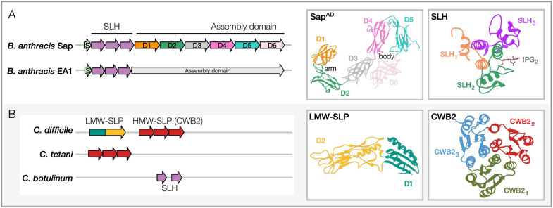
S-layers are self-assembled crystalline 2D lattices enclosing the cell envelopes of several bacteria and archaea. Despite their abundance, the landscape of S-layer structure and function remains a land of wonder. By virtue of their location, bacterial S-layers have been hypothesized to add structural stability to the cell envelope. In addition, S-layers are implicated in mediating cell-environment and cell-host interactions playing a key role in adhesion, cell growth, and division. Significant strides in the understanding of these bacterial cell envelope components were made possible by recent studies that have provided structural and functional insights on the critical S-layer and S-layer-associated proteins (SLPs and SLAPs), highlighting their roles in pathogenicity and their potential as therapeutic or vaccine targets. In this mini-review, we revisit the sequence-structure-function relationships of S-layers, SLPs, and SLAPs in Gram-positive pathogens, focusing on the best-studied classes, Bacilli (Bacillus anthracis) and Clostridia (Clostridioides difficile). We delineate the domains and their architectures in archetypal S-layer proteins across Gram-positive genera and reconcile them with experimental findings. Similarly, we highlight a few key "flavors" of SLPs displayed by Gram-positive pathogens to assemble and support the bacterial S-layers. Together, these findings indicate that S-layers are excellent candidates for translational research (developing diagnostics, antibacterial therapeutics, and vaccines) since they display the three crucial characteristics: accessible location at the cell surface, abundance, and unique lineage-specific signatures.

Keywords: Firmicutes; S-layer; cell envelope; cell surface proteins; gram-positive bacteria; molecular evolution; pathogenicity; sequence-structure features.


Conflict of interest statement

The authors declare that the research was conducted in the absence of any commercial or financial relationships that could be construed as a potential conflict of interest.

Figures

FIGURE 1
FIGURE 1
Sequence-structure features of pathogenic Bacilli and Clostridia SLPs. (A) Bacillus anthracis SLPs, Sap and EA1. Domain architectures: SLH region (containing three SLH domain motifs, Pfam PF00395, ProSiteProfiles PS51272) and the assembly domain are shown for both the representative Bacillus SLPs. PDB structures: Shown for SapAD and Sap-SLH are shown and labeled [PDB: 6HHU, 6BT4 (Kern et al., 2011; Fioravanti et al., 2019)]. The crystal structure of the SLH domain is in complex with a synthetic SCWP unit (IPG). Accession numbers for proteins shown: Sap, AAT53168.1; EA1, AAP24884.1. Similar domain architectures are observed in B. cereus, B. mycoides, and B. thuringiensis SLPs. (B) Clostridia SLPs in Clostridioides difficile, Clostridium tetani, and Clostridium botulinum. Domain architectures: C. difficile contains low-molecular-weight (LMW) and high-molecular-weight (HMW) SLP domains (HMW-SLP with three CWB2 domain motifs; LMW-SLP). C. tetani contains the cell-wall binding domain (CWB2, Pfam PF04122) as well, while C. botulinum carries two SLH domains. PDB structures: Shown for the LMW-SLP and CWB2 of Cwp8 are shown [PDB: 3CVZ, 5J6Q; (Fagan et al., 2009; Usenik et al., 2017)]. Accession numbers for proteins shown: C. difficile, WP_078051019.1; C. tetani, WP_035111087.1; C. botulinum, WP_039307708.1. Domain architectures are marked from cited literature and InterProScan predictions (Jones et al., 2014). Representative PDB structures for Bacillus and Clostridia SLP domains have been redrawn using Phyre2 (Kelley et al., 2015).
FIGURE 2
FIGURE 2
Sequence features of SLPs in key Gram-positive pathogenic genera. Previously documented or predicted [using InterProScan (Jones et al., 2014)] sequence-structure features in representative SLPs from select pathogens in Firmicutes and Actinobacteria. Species considered: B. anthracis, B. cereus, B. mycoides, B. thuringiensis, Clostrioides difficile, Clostridium tetani, Clostridium botulinum, P. alvei, Listeria monocytogenes, L. seeligeri, L. booriae, L. fleischmannii, Streptococcus pneumoniae, S. dysgalactiae, S. pyogenes, Staphylococcus cohnii, S. haemolyticus, Corynebacterium glutamicum, C. aurimucosum, C. minutissimum, M. intracellulare, and M. kansasii. Abbreviations: SLH, S-layer homology domain (Pfam PF00395, ProSiteProfiles PS51272); LMW-SLP, Low molecular weight S layer protein N-terminal (Pfam PF12211); CWB2, Cell wall binding domain 2 (High molecular weight; Pfam PF04122); Extracellular, Region of a membrane-bound protein predicted to be outside the membrane in the extracellular region (Phobius prediction); Outer-MP, Outer membrane protein alpha-related (PANTHER PTHR43308); Bac-Ig-like, Bacterial Ig-like domain (Pfam clan CL0159: PF02368, PF13205); Lyase, Hyaluronate lyase/Polysaccharide lyase family 8 (Pfam PF02278, PF02884, PF08124, PANTHER PTHR38481); Fibronectin, Fibronectin type-III domain (Pfam PS50853); NAM_LA_amidase, N-acetylmuramoyl-L-alanine amidase-related domain (PANTHER PTHR30032); Glucosaminidase, Mannosyl-glycoprotein endo-beta-N-acetylglucosaminidase, Transglutaminase-like superfamily (Pfam PF01832, PF01841); Mem_lipoprot_site, Prokaryotic membrane lipoprotein lipid attachment (ProSiteProfiles PS51257); Glutamine aminotransferase, Glutamine amidotransferase type 2; Glucosamine-fructose-6-phosphate aminotransferase, isomerizing (Pfam PF13522, ProSiteProfiles: PS51278, PANTHER PTHR10937); and Endotoxin, Delta endotoxin (Pfam PF18449).

Similar articles

Cited by

References

    1. Albers S. V., Meyer B. H. (2011). The archaeal cell envelope. Nat. Rev. Microbiol. 9 414–426. 10.1038/nrmicro2576 - DOI - PubMed
    1. Åvall-Jääskeläinen S., Hynönen U., Ilk N., Pum D., Sleytr U. B., Palva A. (2008). Identification and characterization of domains responsible for self-assembly and cell wall binding of the surface layer protein of Lactobacillus brevisATCC 8287. BMC Microbiol. 8:165. 10.1186/1471-2180-8-165 - DOI - PMC - PubMed
    1. Bahl H., Scholz H., Bayan N., Chami M., Leblon G., Gulik-Krzywicki T., et al. (1997). IV. Molecular biology of S-layers. FEMS Microbiol. Rev. 20 47–98. 10.1016/s0168-6445(97)00050-8 - DOI - PubMed
    1. Baillie L., Hebdon R., Flick-Smith H., Williamson D. (2003). Characterisation of the immune response to the UK human anthrax vaccine. FEMS Immunol. Med. Microbiol. 36 83–86. 10.1016/s0928-8244(03)00085-3 - DOI - PubMed
    1. Banerji O., Lanzoni-Mangutchi P., Vaz F., Barwinska-Sendra A., Wilson J., Kirk J., et al. (2021). Structure and assembly of the S-layer determine virulence in C. difficile. Res. Sq. 10.21203/rs.3.rs-79088/v1 - DOI - PMC - PubMed

LinkOut - more resources