

Primary malignant vascular tumors of the liver in children: Angiosarcoma and epithelioid hemangioendothelioma
- PMID: 33889274
- PMCID: PMC8040065
- DOI: 10.4251/wjgo.v13.i4.223
Primary malignant vascular tumors of the liver in children: Angiosarcoma and epithelioid hemangioendothelioma
Abstract
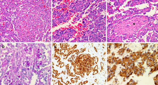
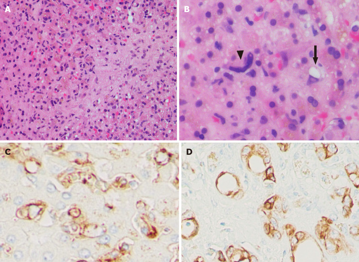
Primary malignant vascular neoplasms of the liver, angiosarcoma and epithelioid hemangioendothelioma, are extremely rare entities in the pediatric population. International Society for the Study of Vascular Anomalies classification system is recommended for the pathologic diagnosis of hepatic vascular lesions in this age group. In this article, we highlight the clinicopathologic characteristics of hepatic angiosarcoma and epithelioid hemangioendothelioma in the pediatric population. Hepatic angiosarcoma in children shows a slight female predominance with an average age of 40 mo at diagnosis. The distinct histologic features include whorls of atypical spindled cells and eosinophilic globules, in addition to the general findings of angiosarcoma. Histologic diagnosis of pediatric hepatic angiosarcoma is not always straightforward, and the diagnostic challenges are discussed in the article. Hepatic epithelioid hemangioendothelioma also demonstrates a female predominance, but is more commonly identified in adolescents (median age at diagnosis: 12 years). Histologically, the lesion is characterized by epithelioid cells and occasional intracytoplasmic lumina with a background of fibromyxoid stroma. While WWTR1-CAMTA1 and YAP1-TFE3 fusions have been associated with epithelioid hemangioendothelioma, there are currently no known signature genetic alterations seen in pediatric hepatic angiosarcoma. Advancement in molecular pathology, particularly for pediatric hepatic angiosarcoma, is necessary for a better understanding of the disease biology, diagnosis, and development of targeted therapies.
Keywords: Epithelioid hemangioendothelioma; Hepatic angiosarcoma; Infantile hemangioma; Liver tumor; Molecular genetics; Pediatric.
©The Author(s) 2021. Published by Baishideng Publishing Group Inc. All rights reserved.
Conflict of interest statement
Conflict-of-interest statement: The authors have nothing to disclose.
Figures
References
-
- Finegold MJ, Egler RA, Goss JA, Guillerman RP, Karpen SJ, Krishnamurthy R, O’Mahony CA. Liver tumors: Pediatric population. Liver Transplant . 2008;14:1545–1556. - PubMed
-
- Chavhan GB, Siddiqui I, Ingley KM, Gupta AA. Rare malignant liver tumors in children. Pediatr Radiol. 2019;49:1404–1421. - PubMed
-
- McGuire A, Fernandez-Pineda I, Fishman SJ, Dickie BH. Pediatric hepatic vascular tumors. Seminars Pediatric Surg. 2020;29:150970. - PubMed
-
- Wassef M, Blei F, Adams D, Alomari A, Baselga E, Berenstein A, Burrows P, Frieden IJ, Garzon MC, Lopez-Gutierrez JC, Lord DJ, Mitchel S, Powell J, Prendiville J, Vikkula M ISSVA Board and Scientific Committee. Vascular Anomalies Classification: Recommendations From the International Society for the Study of Vascular Anomalies. Pediatrics. 2015;136:e203–e214. - PubMed
-
- Christison-Lagay ER, Burrows PE, Alomari A, Dubois J, Kozakewich HP, Lane TS, Paltiel HJ, Klement G, Mulliken JB, Fishman SJ. Hepatic hemangiomas: subtype classification and development of a clinical practice algorithm and registry. J Pediatr Surg. 2007;42:62–7; discussion 67. - PubMed
Publication types
LinkOut - more resources
Full Text Sources
Other Literature Sources
