Review
. 2021 Apr 15;12(4):437-452.
doi: 10.4239/wjd.v12.i4.437.

Malfunction of outer retinal barrier and choroid in the occurrence and progression of diabetic macular edema

Affiliations
Review

Malfunction of outer retinal barrier and choroid in the occurrence and progression of diabetic macular edema

Ştefan Ţălu et al. World J Diabetes. .

Abstract

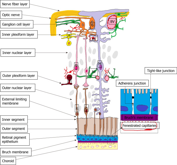
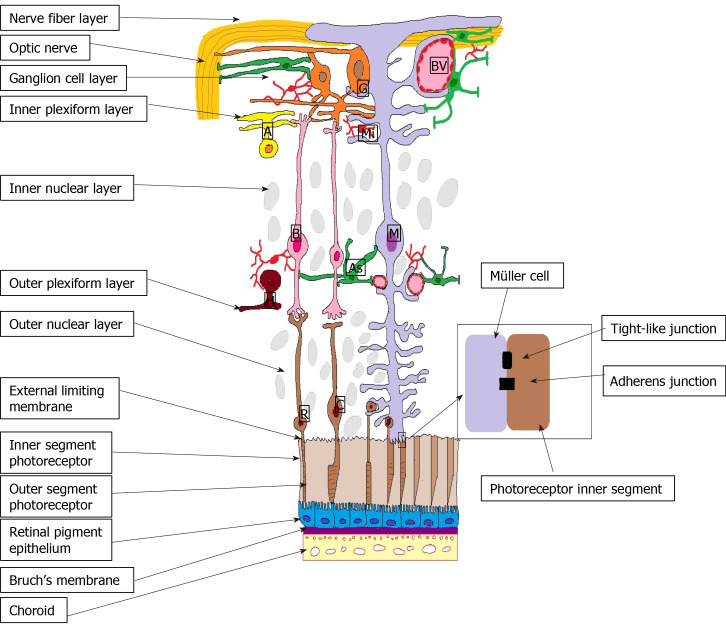
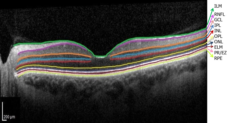
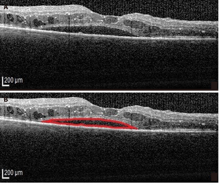
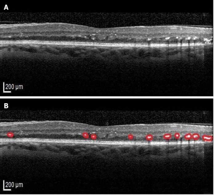
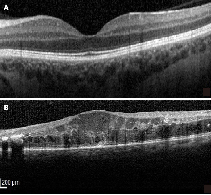
Diabetic macular edema (DME) is the most common cause of vision loss in diabetic retinopathy, affecting 1 in 15 patients with diabetes mellitus (DM). The disruption of the inner blood-retina barrier (BRB) has been largely investigated and attributed the primary role in the pathogenesis and progression in DME, but there is increasing evidence regarding the role of outer BRB, separating the RPE from the underlying choriocapillaris, in the occurrence and evolution of DME. The development of novel imaging technologies has led to major improvement in the field of in vivo structural analysis of the macula allowing us to delve deeper into the pathogenesis of DME and expanding our vision regarding this condition. In this review we gathered the results of studies that investigated specific outer BRB optical coherence tomography parameters in patients with DM with the aim to outline the current status of its role in the pathogenesis and progression of DME and identify new research pathways contributing to the advancement of knowledge in the understanding of this condition.

Keywords: Diabetic macular edema; External limiting membrane; Hyperreflective foci; Inner segment/outer segment line; Optical coherence tomography; Outer retinal barrier.


Conflict of interest statement

Conflict-of-interest statement: The authors declare no conflict of interest regarding the publication of this paper.

Figures

Figure 1
Figure 1
Outer blood-retina barrier. M: Müller cells; A: Amacrine cells; G: Ganglion cells; BV: Blood vessels; As: Astrocyte; B: Bipolar cells; R: Rods; C: Cones; H: Horizontal cells; Mi: Microglia; ON: Optic nerve; NFL: Nerve fibre layer; GCL: Ganglion cell layer; IPL: Inner plexiform layer; INL: Inner nuclear layer; OPL: Outer plexiform layer; ONL: Outer nuclear layer; ELM: External limiting membrane; IS: Inner segment photoreceptors; OS: Outer segment photoreceptors; RPE: Retinal pigment epithelium; BM: Bruch’s Membrane; Ch: Choroid.
Figure 2
Figure 2
External limiting membrane. M: Müller cells; A: Amacrine cells; G: Ganglion cells; BV: Blood vessels; As: Astrocyte; B: Bipolar cells; R: Rods; C: Cones; H: Horizontal cells; Mi: Microglia; ON: Optic nerve; NFL: Nerve fibre layer; GCL: Ganglion cell layer; IPL: Inner plexiform layer; INL: Inner nuclear layer; OPL: Outer plexiform layer; ONL: Outer nuclear layer; ELM: External limiting membrane; IS: Inner segment photoreceptors; OS: Outer segment photoreceptors; RPE: Retinal pigment epithelium; BM: Bruch’s Membrane; Ch: Choroid.
Figure 3
Figure 3
Normal optical coherence tomography aspect of the retinal layers. Segmentation software automatically marked the 10 retinal layers. (ILM: Internal limiting membrane; RNFL: Retinal nerve fiber layer; GCL: Ganglion cell layer; IPL: Inner plexiform layer; INL: Inner nuclear layer; OPL: Outer plexiform layer; ONL: Outer nuclear layer; ELM: External limiting membrane; PR/EZ: Photoreceptor layer/ellipsoid zone (inner and outer photoreceptor segment junction; RPE: Retinal pigment epithelium).
Figure 4
Figure 4
Serous retinal detachment type of diabetic macular edema. A: Optical coherence tomography (OCT)-Retinal neurosensory detachment; B: Highlighted OCT image showing the neurosensory detachment (red).
Figure 5
Figure 5
Hyperreflective foci. A: Original optical coherence tomography (OCT) image; B: Highlighted OCT image revealing hyperreflective foci (red).
Figure 6
Figure 6
Disorganization of the inner retinal layers. A: Normal optical coherence tomography aspect of the macula; B: Disorganization of the inner retinal layers.

References

    1. Kang K, Lee H, Jang M, Kim HC, Chung H. Diabetic macular edema with pachychoroid features. BMC Ophthalmol. 2020;20:392. - PMC - PubMed
    1. Damian I, Nicoara SD. Optical Coherence Tomography Biomarkers of the Outer Blood-Retina Barrier in Patients with Diabetic Macular Oedema. J Diabetes Res. 2020;2020:8880586. - PMC - PubMed
    1. Wong WM, Chee C, Bhargava M, Chai C, Lin H, Zhao P, Ariadarma Mangunkusumo E, Naing T, Yuen YS, Wong TY, Su X, Lingam G. Systemic Factors Associated with Treatment Response in Diabetic Macular Edema. J Ophthalmol. 2020;2020:1875860. - PMC - PubMed
    1. Xu HZ, Le YZ. Significance of outer blood-retina barrier breakdown in diabetes and ischemia. Invest Ophthalmol Vis Sci. 2011;52:2160–2164. - PMC - PubMed
    1. Xia T, Rizzolo LJ. Effects of diabetic retinopathy on the barrier functions of the retinal pigment epithelium. Vision Res. 2017;139:72–81. - PubMed

LinkOut - more resources