. 2021 Apr 13;12(8):783-790.
doi: 10.18632/oncotarget.27941.

Urine protein biomarkers of bladder cancer arising from 16-plex antibody-based screens

Affiliations

Urine protein biomarkers of bladder cancer arising from 16-plex antibody-based screens

Kamala Vanarsa et al. Oncotarget. .

Abstract

Purpose: The purpose of this study is to identify novel urine protein biomarkers of bladder cancer using a Luminex based screening platform.

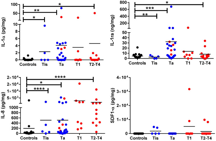
Materials and methods: The current study examines urine samples from 66 subjects, comprised of 31 Urology clinic controls and 35 bladder cancer patients, using a Luminex based screening platform. ELISA validation was carried out for the top 4 prospective urine biomarkers using an independent cohort of 20 Urology clinic controls and 60 bladder cancer (BC) subjects.

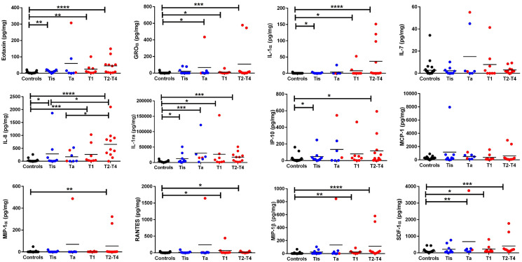
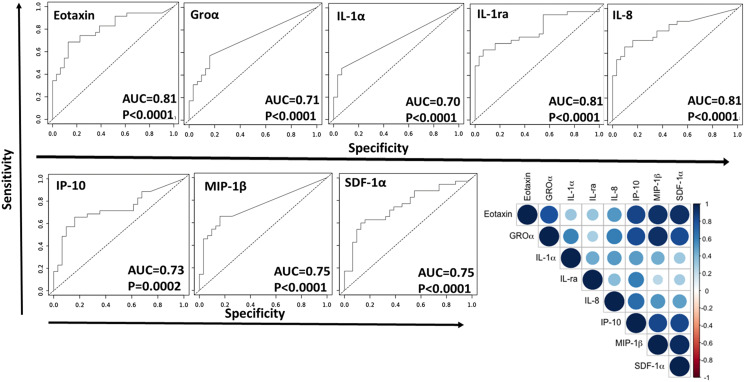
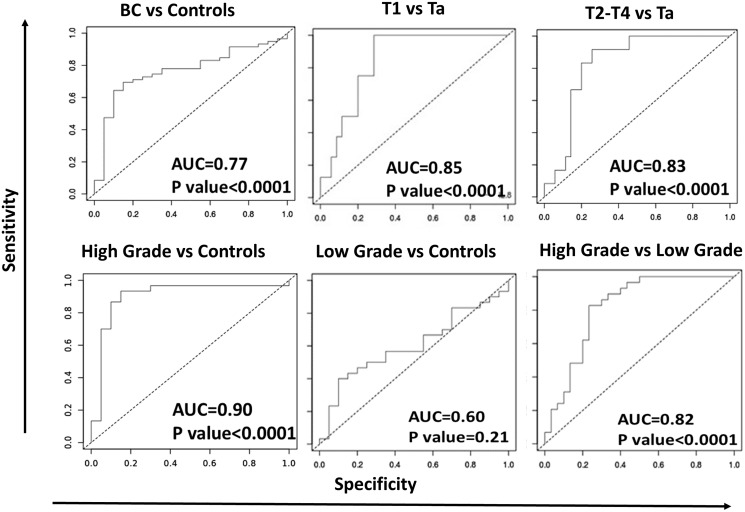
Results: Of the 16 proteins screened by Luminex, 10 showed significant elevation in BC compared to the controls. Eight of these urine proteins were able to differentiate BC from control urine with ROC AUC values exceeding 0.70 at p < 0.0001, with specificity values exceeding 0.9. Upon ELISA validation, urine IL-1α, IL-1ra, and IL-8 were able to distinguish control urine from urine drawn from various bladder cancer stages, with IL-8 being the best discriminator. Compared to members of the IL-1 cytokine family, urine IL-8 was also best at discriminating T1 and/or T2-T4 from Ta BC (ROC AUC ≥ 0.83), as well as high grade from low grade BC (ROC AUC ≥ 0.82).

Conclusions: These findings suggest that urine IL-1α, IL-1ra and IL-8 are useful indicators of bladder cancer. Urine IL-8 not only distinguishes bladder cancer from controls, it also discriminates high grade from low grade disease, and the successive clinical stages of bladder cancer. While supportive of previous reports, these findings warrant further analysis in prospective cohorts.

Keywords: inflammation; interleukins; proteomics; targeted screens; urothelial.


Conflict of interest statement

CONFLICTS OF INTEREST Authors have no conflicts of interest to declare.

Figures

Figure 1
Figure 1. Luminex based screen of 16 urine proteins in bladder cancer.
The dot plots depict the 12 proteins that were detectable by Luminex assay either in BC urine or the urology clinic controls, labeled as “Controls”. Tested samples included 31 controls, 7 Ta, 9 Tis, 8 T1, and 11 T2–T4 urine samples. Creatinine normalized urine protein levels are shown in different colors specific for each group (black dots = controls, blue dots = low grade BC, and red dots = high grade BC). Low grade tumor and high grade tumor classification was based on pathology reports. The asterisks designate the level of significance between the different groups: * = p < 0.05, ** = p < 0.01, *** = p < 0.001, and **** = p < 0.0001, using a Mann Whitney U test. The primary data for this analysis is presented in Supplementary Table 2. All controls used for this study were drawn from the Urology clinic, including patients investigated for hematuria but found not to have any urological cancers.
Figure 2
Figure 2. Eight urine proteins that discriminate BC from controls based on the Luminex-based screen of 16 proteins.
Receiver Operating Curve Area Under Curve (ROC-AUC) plots were generated for eight urine proteins including Eotaxin, GROα, IL-1α, IL-1ra, IL-8, IP-10, MIP-1β, and SDF-1α to determine their ability to discriminate BC from controls. AUC values and p-values are listed on each curve. The closer the AUC value is to 1, the higher the discriminatory potential of the protein to distinguish between the two groups, with maximized specificity and sensitivity. All of the proteins exhibited AUC values of 0.70 or higher, with p-values < 0.0001, except IP-10 which had a p-value of 0.0002. A correlation plot was also generated for these eight urine proteins. Each circle represents the degree of correlation for the given protein pair, with blue intensity corresponding to positive correlation and red intensity corresponding to negative correlation.
Figure 3
Figure 3. ELISA validation of IL-1α, IL-1ra, IL-8, and SDF-1α in bladder cancer patients with varying clinical stages.
The dot plots depict the expression of IL-1α, IL-1ra, IL-8, and SDF-1α in urine from different stages of BC. Included were 20 urology clinic controls (“controls”), 35 Ta, 5 Tis, 8 T1, and 12 T2-T4 BC patients. Creatinine normalized urine protein levels are shown in different colors (black dots = controls, blue dots = low grade BC and red dots = high grade BC). Low grade tumor and high grade tumor classification was based on pathology reports. The asterisks designate the level of significance between the different groups: *= p < 0.05, **= p < 0.01, ***= p < 0.001, and **** = p < 0.0001, using a Mann Whitney U test.
Figure 4
Figure 4. ROC-AUC curves for urine IL-8 in distinguishing different stages of bladder cancer.
ROC-AUC curves were generated for urine IL-8 to determine its discriminatory capability among different BC groups. AUC values and p-values are listed on each curve. The closer the AUC value is to 1, the higher the discriminatory potential of the protein to distinguish between the two sample groups, with maximized sensitivity and specificity. All comparisons exhibited AUC values of 0.77 or higher, with p-values < 0.0001, except for the comparison between low grade versus urology clinic controls.

References

    1. Siegel RL, Miller KD, Jemal A. Cancer statistics, 2020. CA Cancer J Clin. 2020; 70:7–30. 10.3322/caac.21590. - DOI - PubMed
    1. Cancer Facts & Figures 2020. American Cancer Society. https://www.cancer.org/research/cancer-facts-statistics/.
    1. Saginala K, Barsouk A, Aluru JS, Rawla P, Padala SA, Barsouk A. Epidemiology of Bladder Cancer. Med Sci (Basel). 2020; 8:15. 10.3390/medsci8010015. - DOI - PMC - PubMed
    1. Chakraborty A, Dasari S, Long W, Mohan C. Urine protein biomarkers for the detection, surveillance, and treatment response prediction of bladder cancer. Am J Cancer Res. 2019; 9:1104–1117. - PMC - PubMed
    1. Soria F, Droller MJ, Lotan Y, Gontero P, D'Andrea D, Gust KM, Rouprêt M, Babjuk M, Palou J, Shariat SF. An up-to-date catalog of available urinary biomarkers for the surveillance of non-muscle invasive bladder cancer. World J Urol. 2018; 36:1981–1995. 10.1007/s00345-018-2380-x. - DOI - PMC - PubMed

LinkOut - more resources