Review
. 2021 Mar 6:10:100107.
doi: 10.1016/j.mtbio.2021.100107. eCollection 2021 Mar.

Scaffold-based developmental tissue engineering strategies for ectodermal organ regeneration

Affiliations
Review

Scaffold-based developmental tissue engineering strategies for ectodermal organ regeneration

N Contessi Negrini et al. Mater Today Bio. .

Abstract

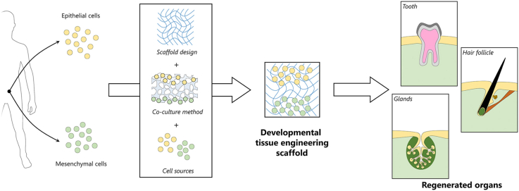
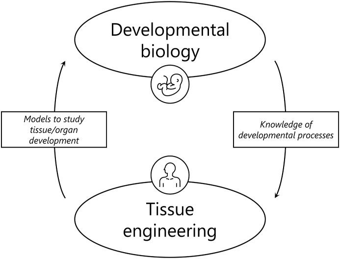
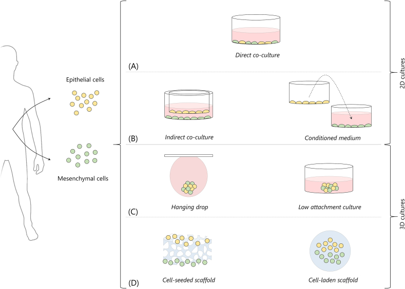
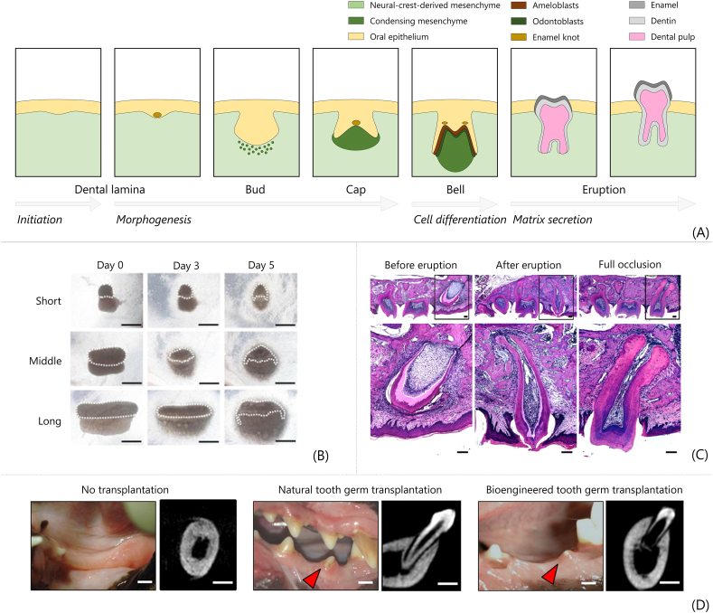
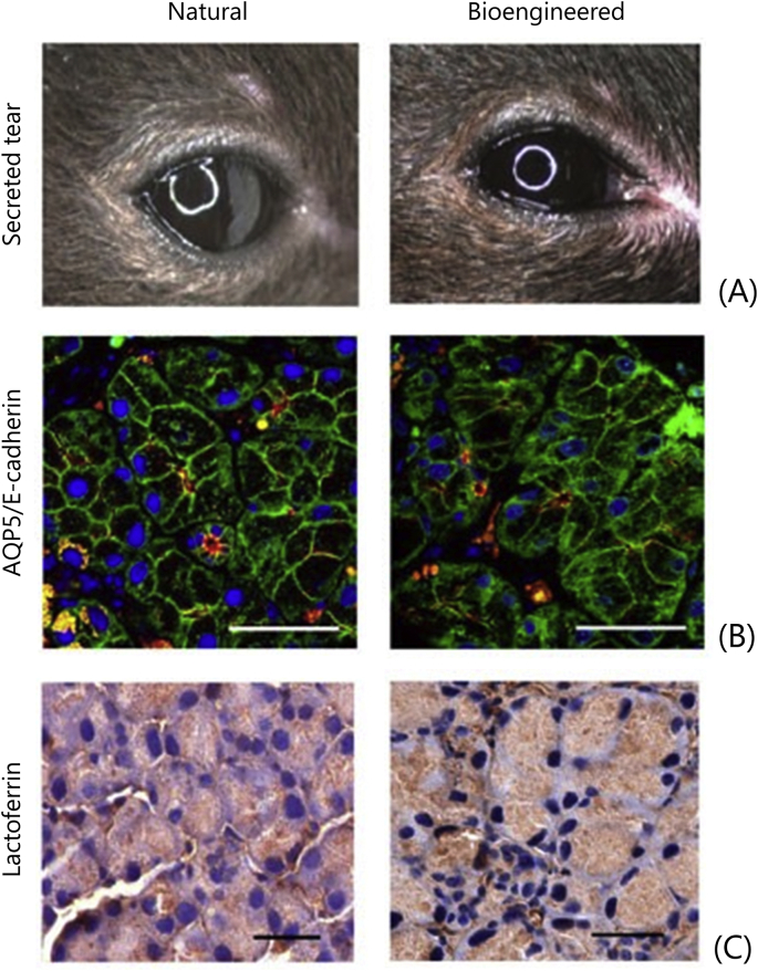
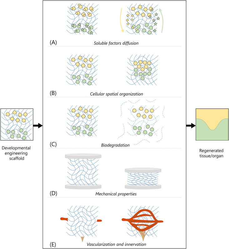
Tissue engineering (TE) is a multidisciplinary research field aiming at the regeneration, restoration, or replacement of damaged tissues and organs. Classical TE approaches combine scaffolds, cells and soluble factors to fabricate constructs mimicking the native tissue to be regenerated. However, to date, limited success in clinical translations has been achieved by classical TE approaches, because of the lack of satisfactory biomorphological and biofunctional features of the obtained constructs. Developmental TE has emerged as a novel TE paradigm to obtain tissues and organs with correct biomorphology and biofunctionality by mimicking the morphogenetic processes leading to the tissue/organ generation in the embryo. Ectodermal appendages, for instance, develop in vivo by sequential interactions between epithelium and mesenchyme, in a process known as secondary induction. A fine artificial replication of these complex interactions can potentially lead to the fabrication of the tissues/organs to be regenerated. Successful developmental TE applications have been reported, in vitro and in vivo, for ectodermal appendages such as teeth, hair follicles and glands. Developmental TE strategies require an accurate selection of cell sources, scaffolds and cell culture configurations to allow for the correct replication of the in vivo morphogenetic cues. Herein, we describe and discuss the emergence of this TE paradigm by reviewing the achievements obtained so far in developmental TE 3D scaffolds for teeth, hair follicles, and salivary and lacrimal glands, with particular focus on the selection of biomaterials and cell culture configurations.

Keywords: Cell coculture; Developmental; Epithelial-mesenchymal interaction; Gland regeneration; Hair follicle regeneration; Tooth regeneration; tissue engineering.


Conflict of interest statement

The authors declare that they have no known competing financial interests or personal relationships that could have appeared to influence the work reported in this article.

Figures

Image 1
Graphical abstract
Fig. 1
Fig. 1
Interplay between developmental biology and tissue engineering. Developmental biology provides knowledge on biological developmental processes to be used for human tissue regeneration by tissue engineering, while tissue engineering provides models to investigate developmental processes in developmental biology research (icons © The Noun Project).
Fig. 2
Fig. 2
Strategies for cell cocultures to replicate developmental processes. 2D cocultures are based on (A) direct and (B) indirect cocultures. Indirect cocultures can be performed by transwell culture systems (left) or by culturing a cell population with a conditioned medium used to previously culture the other cell population (right). 3D cocultures can be based on (C) scaffold-free approaches, such as hanging drop method (left) or ultra-low attachment cultures (right) or (D) scaffold-based approaches, including cell seeding on scaffolds (left) or cells embedding in scaffolds, typically hydrogel-based (right). Culture medium is represented in red; biomaterials/scaffolds are represented in blue.
Fig. 3
Fig. 3
Tooth developmental tissue engineering. (A) Fundamental steps of tooth morphogenesis in embryos replicated by developmental TE to regenerate teeth. (B) Phase contrast images of bioengineered tooth germs in collagen drops (the ‘collagen drop organ germ method’). Epithelial (dashed lines) and mesenchymal cells are seeded compartmentalized in collagen drops with different contact areas (short, middle and long); scale bar ​= ​200 ​μm [99]. (C) Histological sections of bioengineered tooth fabricated by collagen drop method implanted in mice upper first molar, before and immediately after eruption, and at complete occlusion; scale bar ​= ​100 ​μm [100]. (D) Macroscopic pictures and CT images of canine oral cavity without transplantation, after natural tooth germ transplantation, and after in vitro bioengineering tooth transplantation. Red arrows indicate erupted tooth [101]. All images used with permission.
Fig. 4
Fig. 4
Schematic (i) and fluorescent image (ii, scale bar 1 ​mm) of coculture dental epithelial cells (green) and dental mesenchymal cells. Confocal image of the area at the interface between the hydrogels (iii, scale bar 100 ​μm) and magnification of the area in the white box showing polarized cells (iv, scale bar 20 ​μm) [122]. All images used with permission.
Fig. 5
Fig. 5
Hair follicle developmental tissue engineering. (A) Fundamental steps of hair follicle morphogenesis in embryos replicated by developmental TE. (B) Histological sections of intracutaneously regenerated hair follicle. Haematoxylin and eosin staining (H&E, upper row) and green fluorescent protein (GFP)/nuclei fluorescent images. Panels on the right show higher magnifications of the boxed are in the figures on the left (scale bar 100 ​μm) [152]. (C) Integration of transplanted HFs with surrounding tissues. Black hair shafts regenerated among native white hair shafts of nude mice. Calponin staining in red for muscle (i.e. arrector pili muscles), neurofilament H staining in white for nerve fibres, and nuclei in blue; scale bar 200 ​μm for lower magnification and 100 ​μm for higher magnification (reprinted/adapted from Ref. [153]; © The Authors, some rights reserved; exclusive licensee American Association for the Advancement of Science. Distributed under a Creative Commons Attribution Non-Commercial License 4.0 (CC BY-NC) http://creativecommons.org/licenses/by-nc/4.0/). (D) Morphological images of bioengineered hair follicle cycle in vivo over 80 days period (scale bar 1 ​mm) [154]. All images used with permission.
Fig. 6
Fig. 6
Fabrication and transplantation of spatially aligned hair follicle germs. (A) Germs are generated by coculturing cells in non-adhesive wells. Then, a collagen gel with a support mesh is used to embed spatially aligned germs and the obtained collagen sheet is used for transplantation. (B) Phase-contrast (scale bar ​= ​1 ​mm) and H&E staining images (scale bar ​= ​200 ​μm) of cell cocultures performed in oxygen-permeable wells PDMS (left) and non-oxygen-permeable polymethyl methacrylate (PMMA) wells (right). (C) Regenerated aligned hair follicles after 18 days of transplantation in back skin of nude mice [167]. All images used with permission.
Fig. 7
Fig. 7
Salivary gland developmental tissue engineering. (A) Fundamental steps of salivary gland in vivo morphogenesis. (B) Phase contrast images and H&E staining of natural and bioengineered salivary glands, obtained by developmental TE, cultured in vitro up to 3 days; scale bar ​= ​200 ​μm [173]. All images used with permission.
Fig. 8
Fig. 8
Lacrimal gland developmental tissue engineering. (A) Tear secretion from mice with natural lacrimal gland (left) and bioengineered lacrimal gland (right). (B) Immunohistochemical images of acinar and duct cells in natural and bioengineered lacrimal glands stained for aquaporin-5 (AQP5, red) and E-cadherin (green); scale bar ​= ​50 ​μm. (C) Lactoferrin in the acini of natural and bioengineered implanted lacrimal gland; scale bar ​= ​50 ​μm [174]. All images used with permission.
Fig. 9
Fig. 9
Developmental tissue engineering scaffold requirements. (A) Diffusion of nutrients and oxygen for cell survival as well as soluble factors fundamental for epithelial-mesenchymal interactions. (B) Possibility for cells to remodel the artificial ECM and correctly organize in space to develop the regenerated tissue/organ. (C) Biodegradability of the scaffold that should be, in time, substituted with the newly formed ECM of the mature tissue/organ. (D) Adequate mechanical properties. (E) Promotion of vascularization and innervation to allow for the mature tissue/organ survival and functionality. Epithelial and mesenchymal cells are depicted in yellow and green; light blue lines represent a generic 3D developmental tissue engineering scaffold.

References

    1. Langer R., Vacanti J.P. Tissue engineering. Science. 1993;260:920–926. doi: 10.1126/science.8493529. (80-.) - DOI - PubMed
    1. Kim Y.S., Smoak M.M., Melchiorri A.J., Mikos A.G. An overview of the tissue engineering market in the United States from 2011 to 2018. Tissue Eng. 2019;25:1–8. doi: 10.1089/ten.tea.2018.0138. - DOI - PMC - PubMed
    1. Caddeo S., Boffito M., Sartori S. Tissue engineering approaches in the design of healthy and pathological in vitro tissue models. Front. Bioeng. Biotechnol. 2017;5:1–22. doi: 10.3389/fbioe.2017.00040. - DOI - PMC - PubMed
    1. Takahashi K., Yamanaka S. Induction of pluripotent stem cells from mouse embryonic and adult fibroblast cultures by defined factors. Cell. 2006;126:663–676. doi: 10.1016/j.cell.2006.07.024. - DOI - PubMed
    1. Khademhosseini A., Langer R. A decade of progress in tissue engineering. Nat. Protoc. 2016;11:1775–1781. doi: 10.1038/nprot.2016.123. - DOI - PubMed

LinkOut - more resources