Review
. 2021 Aug 17;144(7):1958-1973.
doi: 10.1093/brain/awab059.

The extracellular matrix as modifier of neuroinflammation and remyelination in multiple sclerosis

Affiliations
Review

The extracellular matrix as modifier of neuroinflammation and remyelination in multiple sclerosis

Samira Ghorbani et al. Brain. .

Abstract

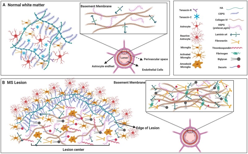
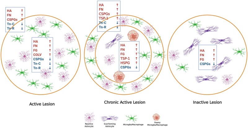
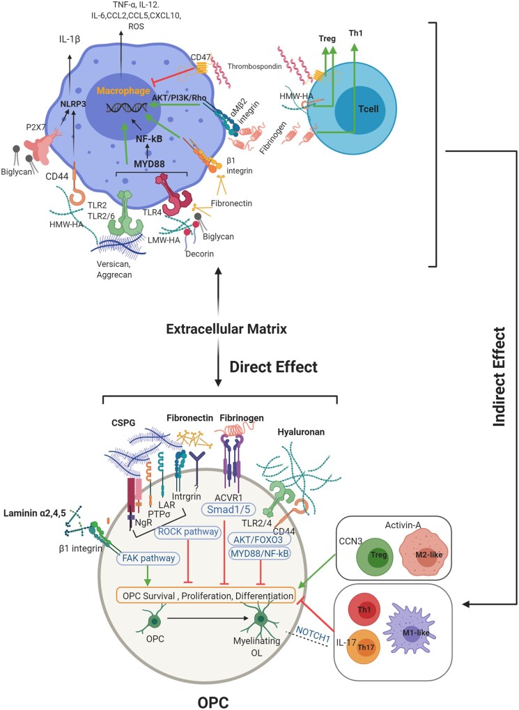
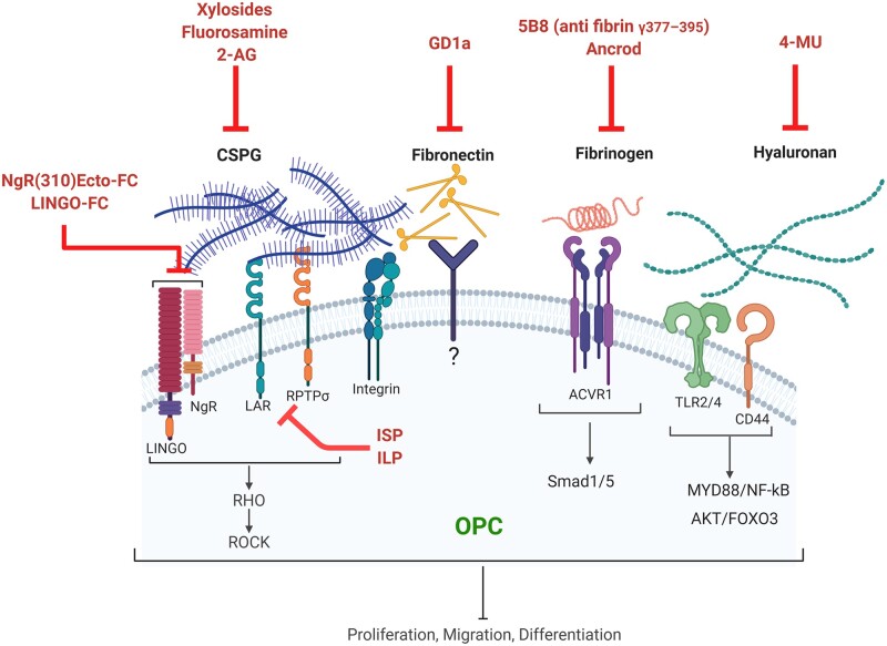
Remyelination failure contributes to axonal loss and progression of disability in multiple sclerosis. The failed repair process could be due to ongoing toxic neuroinflammation and to an inhibitory lesion microenvironment that prevents recruitment and/or differentiation of oligodendrocyte progenitor cells into myelin-forming oligodendrocytes. The extracellular matrix molecules deposited into lesions provide both an altered microenvironment that inhibits oligodendrocyte progenitor cells, and a fuel that exacerbates inflammatory responses within lesions. In this review, we discuss the extracellular matrix and where its molecules are normally distributed in an uninjured adult brain, specifically at the basement membranes of cerebral vessels, in perineuronal nets that surround the soma of certain populations of neurons, and in interstitial matrix between neural cells. We then highlight the deposition of different extracellular matrix members in multiple sclerosis lesions, including chondroitin sulphate proteoglycans, collagens, laminins, fibronectin, fibrinogen, thrombospondin and others. We consider reasons behind changes in extracellular matrix components in multiple sclerosis lesions, mainly due to deposition by cells such as reactive astrocytes and microglia/macrophages. We next discuss the consequences of an altered extracellular matrix in multiple sclerosis lesions. Besides impairing oligodendrocyte recruitment, many of the extracellular matrix components elevated in multiple sclerosis lesions are pro-inflammatory and they enhance inflammatory processes through several mechanisms. However, molecules such as thrombospondin-1 may counter inflammatory processes, and laminins appear to favour repair. Overall, we emphasize the crosstalk between the extracellular matrix, immune responses and remyelination in modulating lesions for recovery or worsening. Finally, we review potential therapeutic approaches to target extracellular matrix components to reduce detrimental neuroinflammation and to promote recruitment and maturation of oligodendrocyte lineage cells to enhance remyelination.

Keywords: CSPGs; extracellular matrix; multiple sclerosis; remyelination.


Figures

Figure 1
Figure 1
The ECM in the healthy CNS and in multiple sclerosis lesion. (A) In the uninjured white matter of the CNS, the neural interstitial matrix in the parenchyma primarily consists of CSPGs, hyaluronan (HA) and tenascins whereas collagens (principally type IV), laminins and some members of HSPGs are concentrated in the basement membranes that separate the perivascular space post-endothelial barrier. (B) Several members of the ECM are altered in multiple sclerosis lesions. High levels of CSPGs and hyaluronan accumulate and are prominent at the hypercellular edge as they have been deposited and then cleared from the centre of chronic active multiple sclerosis lesions. As well, fibrillar collagens (I, III, V), small leucine-rich repeat proteoglycans (biglycan, decorin), thrombospondin and the HSPG member (e.g. perlecan) are upregulated in the parenchyma of lesions. Moreover, the basement membranes in multiple sclerosis show a meshwork of ECM components including increased laminins, HSPGs, fibronectin, biglycan, decorin and collagens. Images were created using BioRender.
Figure 2
Figure 2
Expression of ECM proteins in different white matter multiple sclerosis lesions. Schematic shows ECM changes in distinct multiple sclerosis lesion types including active, chronic active and inactive lesions. Early demyelinating active lesions consist of hypercellular lesion centre containing reactive astrocytes and immune cells. Chronic active lesions are defined by a hypercellular inflammatory margin and a hypocellular centre with fibrous astrocytes and foamy (myelin-laden) microglia/macrophages. Inactive lesions show minimal signs of inflammation while containing mainly scar forming (fibrous) astrocytes. Each lesion type has a different ECM composition when compared among each other and to normal white matter. Refer to the main text for the capacity of individual ECM components to modulate the activity of immune cells. In active lesions, prominent sources of hyaluronan (HA), fibronectin (FN), CSPGs and Tenascin-C/R (Tn-C/R) appear to be reactive astrocytes while fibrinogen (FG) is deposited by leakage of serum into lesions. Sources of CSPGs are reactive astrocytes and macrophages/microglia although these cells are also removing the deposited CSPGs from the lesion centre as the immune cells move outwards. The accumulation of inhibitory ECM such as CSPGs in the lesion edge is thought to be a barrier to incoming progenitor cells such as OPCs that attempt to repair the lesion. COLV = collagen V. Images were created using BioRender.
Figure 3
Figure 3
Direct interaction between ECM molecules and OPCs, and indirect effect of ECM on immune cells, affect OPCS and remyelination. This schematic aims to emphasize that remyelination is an outcome of the direct interplay between OPCs and ECM, and indirectly through the effects of ECM on immune cell activity. While CSPGs, hyaluronan, decorin, biglycan, fibrinogen and fibronectin promote an M1-like phenotype in macrophages, thrombospondin drives a regulatory response in both macrophages and T cells. The different phenotype of macrophages or T cells then contribute to oligodendrocyte development in diverse ways. Not strongly emphasized in this review, but also prominent, is that pro-inflammatory macrophages, Th1 and Th17 cells can directly impede OPCs, while a switch towards a regulatory phenotype such as M2-like macrophage and Treg improves tissue repair. ACVR1 = activin A receptor, type I; CCN3 = cellular communication network factor 3; FAK = focal adhesion kinase; Foxo3 = forkhead box O-3; HA = hyaluronan; HWA-HA = high molecular weight hyaluronan; LMW-HA = low molecular weight hyaluronan; LAR = leucocyte common antigen related; MYD88 = myeloid differentiation primary response 88; NF-kB = nuclear factor kappa-light-chain-enhancer of activated B cells; NgR = Nogo receptor; NLRP3 = NLR family pyrin domain containing 3; NOTCH1 = Notch homolog 1, translocation-associated; PTPσ = protein tyrosine phosphatase sigma; ROCK = Rho-associated protein kinase; ROS = reactive oxygen species; Smad1/5 = small mothers against decapentaplegic 1/5. Images were created using BioRender.
Figure 4
Figure 4
Targeting ECM components to promote OPCs and remyelination. This schematic depicts different approaches to overcome the inhibitory effect of dysregulated ECM molecules in multiple sclerosis. These methods include blocking hyaluronan synthesis by 4-methylumbelliferone (4-MU); blocking CSPGs synthesis by xylosides, fluorosamine (4-fluoro-N-acetylglucosamine) or 2-arachidonoylglycerol (2-AG); interfering with CSPG signalling through PTPσ/LAR-specific blocking peptides (ISP, ILP); antagonizing Nogo receptor complex using blocking antibodies; depleting fibrinogen by ancrod or blocking its activity using an anti-fibrin antibody; and neutralizing aggregated fibronectin by ganglioside GD1a. ACVR1 = activin A receptor, type I; Foxo3 = forkhead box O-3; ILP = intracellular LAR peptide; ISP = intracellular sigma peptide; LAR = leucocyte common antigen related; NgR = Nogo receptor; MYD88 = myeloid differentiation primary response 88; NF-kB = nuclear factor kappa-light-chain-enhancer of activated B cells; PTPσ = protein tyrosine phosphatase Sigma; ROCK = Rho-associated protein kinase; Smad1/5 = small mothers against decapentaplegic1/5. Images were created using BioRender.

References

    1. Croxford AL, Spath S, Becher B.. GM-CSF in neuroinflammation: licensing myeloid cells for tissue damage. Trends Immunol. 2015;36:651–662. - PubMed
    1. Plemel JR, Liu WQ, Yong VW.. Remyelination therapies: a new direction and challenge in multiple sclerosis. Nat Rev Drug Discov. 2017;16:617–634. - PubMed
    1. Lau LW, Cua R, Keough MB, Haylock-Jacobs S, Yong VW.. Pathophysiology of the brain extracellular matrix: a new target for remyelination. Nat Rev Neurosci. 2013;14:722–729. - PubMed
    1. Sorokin L.The impact of the extracellular matrix on inflammation. Nat Rev Immunol. 2010;10:712–723. - PubMed
    1. Rauch U.Brain matrix: structure, turnover and necessity. Biochem Soc Trans. 2007;35:656–660. - PubMed

Publication types

Grants and funding