. 2021 Jun 22;144(5):1590-1602.
doi: 10.1093/brain/awab044.

Spatiotemporal dynamics between interictal epileptiform discharges and ripples during associative memory processing

Affiliations

Spatiotemporal dynamics between interictal epileptiform discharges and ripples during associative memory processing

Simon Henin et al. Brain. .

Abstract

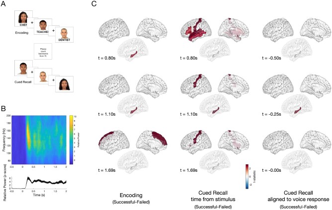
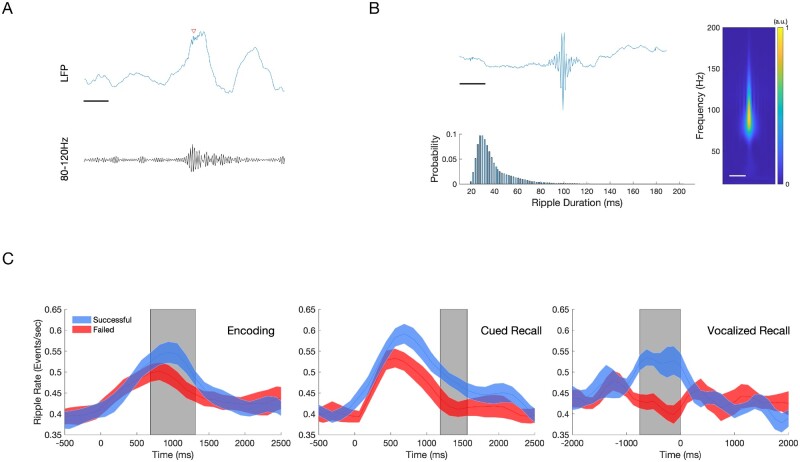
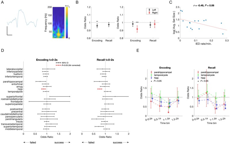
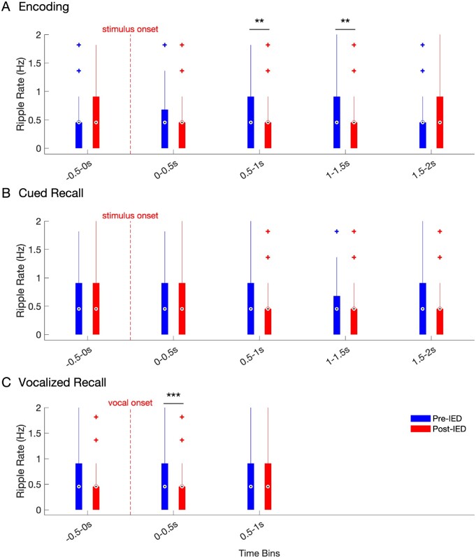
We describe the spatiotemporal course of cortical high-gamma activity, hippocampal ripple activity and interictal epileptiform discharges during an associative memory task in 15 epilepsy patients undergoing invasive EEG. Successful encoding trials manifested significantly greater high-gamma activity in hippocampus and frontal regions. Successful cued recall trials manifested sustained high-gamma activity in hippocampus compared to failed responses. Hippocampal ripple rates were greater during successful encoding and retrieval trials. Interictal epileptiform discharges during encoding were associated with 15% decreased odds of remembering in hippocampus (95% confidence interval 6-23%). Hippocampal interictal epileptiform discharges during retrieval predicted 25% decreased odds of remembering (15-33%). Odds of remembering were reduced by 25-52% if interictal epileptiform discharges occurred during the 500-2000 ms window of encoding or by 41% during retrieval. During encoding and retrieval, hippocampal interictal epileptiform discharges were followed by a transient decrease in ripple rate. We hypothesize that interictal epileptiform discharges impair associative memory in a regionally and temporally specific manner by decreasing physiological hippocampal ripples necessary for effective encoding and recall. Because dynamic memory impairment arises from pathological interictal epileptiform discharge events competing with physiological ripples, interictal epileptiform discharges represent a promising therapeutic target for memory remediation in patients with epilepsy.

Keywords: epilepsy; epileptiform discharges; gamma oscillations; interictal spikes; intracranial EEG.


Figures

Figure 1
Figure 1
Task design and differences in HGA between successful and failed associative memory. (A) A computerized program presented task stimuli and recorded subject spoken responses. During encoding, each face-profession pair was shown for 5 s, with a 1-s interstimulus interval (ISI), which was marked by a plus symbol. To ensure attention and sensory processing of test stimuli, subjects were instructed to read the profession aloud and make a mental association. To prevent rehearsal, a brief distraction task followed the encoding block, during which subjects were asked to count backwards from 15. During cued recall, subjects were shown only the faces from the prior set and asked to say aloud the associated profession. The cued recall period lasted for as long as the subject needed to provide a response. Voice response was recorded for the last 10 subjects and scored for accuracy. (B) Example spectrogram (top) of the raw data recorded in the occipital cortex, and high gamma activity (bottom, HGA 60–170 Hz) normalized to the −500 prestimulus baseline, with a peak at 250 ms after stimulus presentation. (C) Group-level differences in HGA by time for correctly versus incorrectly recalled face-profession pairs thresholded at P < 0.05 (cluster-corrected) during encoding (left), cued recall (middle) and vocal-aligned cued recall (right). Left: During encoding, increased HGA in hippocampus beginning approximately +0.80s after stimulus presentation, with increased HGA in superior frontal region beginning approximately +1.69 s distinguished between successful and failed trials. Middle: During cued recall, increased HGA at +0.80 s after face stimulus presentation in inferior frontal gyrus, postcentral, superior temporal and middle temporal gyrus, and later at +1.10 s in hippocampus distinguish between successful and failed trials P < 0.05, cluster-corrected). Right: To disambiguate the contribution of vocalization to cued recall, the difference between successful and failed trials was determined, timed in response to the vocalization in 10 patients. A difference in hippocampal HGA was seen beginning at −250 ms prior to vocalization (all significant clusters identified at a significance threshold P < 0.05 using a cluster-based permutation test).
Figure 2
Figure 2
Electrode coverage for 15 subjects. We recorded from a total of 1646 electrodes in 15 subjects. Five patients had bilateral depths and strips (Patients NY609, NY639, NY645, NY723, NY736). Six patients had left hemisphere subdural grids, strips, and depths (Patients NY704, NY 708, NY717, NY741, NY743, NY748). Four patients had right hemisphere subdural grids, strips, and depths (Patients NY652, NY661, NY733, NY737). Grid and strip electrodes are shown in red; depth electrodes are shown in blue. Patient NY737 did not have hippocampal depth electrodes and therefore was excluded from ripple analysis.
Figure 3
Figure 3
Hippocampal ripples during encoding and recall predict successful associative memory. Ripple events were detected using a bipolar montage from the electrodes located in or closest to the hippocampus, using a previously published method (80–120 Hz, 20–200 ms duration). To reduce the detections of pathological high frequency oscillations (HFOs), detections were restricted to regions outside of SOZ, and with trials which did not contain an IED. (A) Sample of a raw EEG tracing (blue) with detected ripple event (red arrow), with bandpass filtered (80–120 Hz) tracing (black). Scale bar = 125 ms (B) Characteristics of all detected hippocampal ripples. An average of 1955 ripple events were detected per patient across all conditions. Top: Grand averaged ripple response (left) and spectrogram (right, 10–200 Hz) demonstrates a peak frequency between 80 Hz and 100 Hz. Scale bars = 125 ms. Bottom: Histogram showing detected ripple duration, which follows skewed log distribution. Mean ripple duration 39.8 ms, SD 18.3 ms. (C) Average ripple rate between Successful and Failed Associative Memory Trials (mean ± standard error of the mean). Top: Successful encoding is characterized by a higher hippocampal ripple rate (blue) compared to failed encoding (red) between 750 ms and1375 ms after stimulus presentation (grey box, n =14, P <0.05, cluster-corrected). Middle: Successful cued recall is characterized by a higher ripple rate compared to failed cued recall between +1250 ms and +1625 ms after stimulus presentation (grey box, n =14, P <0.05, cluster-corrected). Bottom: Successful cued recall is characterized by a higher ripple rate from −750 ms to 0 ms aligned voice response (grey box, n =9, P <0.05, cluster-corrected).
Figure 4
Figure 4
IEDs and effect on memory performance. (A) Example raw tracing and spectrogram of a detected IED. Scale bars = 25 µV, 125 ms. (B) Left: IEDs recorded in any brain region during encoding predicted a 4% decreased odds of remembering (OR = 0.96, CI = 0.93–0.99, P =0.002, F-test). IEDs in any brain region during cued recall trended towards decreased odds of remembering (P =0.0760, F-test). Right: IEDs occurring in the right hemisphere predicted a 9% decreased odds of remembering (P =0.0002, F-test). There was no difference in odds of remembering for IEDs in the left hemisphere during encoding, or either hemisphere during recall. Error bars represent 95% confidence intervals. (C) Relationship between block size, IQ, and IED rate. Subjects varied in performance, ranging between 1 and 10 stimuli presented per block, larger sets indicating superior task performance across patients. There was a trend towards a negative correlation between IED rate and log set size [r(13) = −0.49, P =0.06]. (D) Odds of successful memory for IEDs occurring during encoding and recall, by brain region. Mean odds of successful memory per IED occurring during encoding (left) and cued recall (right). Error bars represent 95% CI. Odds < 1 indicate a decreased odds of successful remembering if an IED occurred during the trial. After correction for multiple comparisons, a significant decrease in remembering occurs for IEDs in hippocampus [red, encoding: OR = 0.85, CI = 0.77–0.94), F(1,6221) = 10.5, P =0.001; recall: OR = 0.75, CI = 0.67–0.85, F(1,5956) = 21.8, P <0.001]. (E) Odds of successful memory for IEDs occurring in selected brain regions, by 500 ms time bin. Mean odds of successful memory per IED occurring during encoding (left), demonstrate that odds of remembering are further decreased by 25–52% if IEDs occurred in hippocampus, parahippocampal gyrus, and temporal pole occur between 500–2000 ms. During recall (right), mean odds of successful memory per IED in hippocampus decreased by 41% when IEDs occurred between 1000 ms and 2000 ms after stimulus presentation. Error bars represent 95% CI. Significant changes indicated by an asterisk (P <0.05, F-test).
Figure 5
Figure 5
Hippocampal ripple rate in the pre and post-IED window during encoding and recall. Ripple rates across all hippocampal electrodes in 500 ms pre- (blue) versus 500 ms post-IED (red), binned by time of detected IEDs (500-ms bin windows) for IEDs detected during (A) encoding, (B) cued recall, and (C) voice-aligned recall. Box and whisker plots represent the median (circles) and IQR (bars), along with extreme values (whiskers and outliers). A reduction in ripple rate after an IED event was found during the 0.5–1 s (Z =2.8838, nIEDs = 209, P =0.004) and 1–1.5 s (Z =3.0873, nIEDs=266, P =0.002) windows during encoding. In addition, ripple rates were significantly reduced in the time window during (0–0.5 s) vocalization during cued recall (Z =3.9, nIEDs = 177, P < 0.001).

References

    1. Fisher RS, Vickrey BG, Gibson P, et al.The impact of epilepsy from the patient's perspective I. Descriptions and subjective perceptions. Epilepsy Res. 2000;41:39-51. - PubMed
    1. Lemesle B, Planton M, Pages B, et al.Accelerated long-term forgetting and autobiographical memory disorders in temporal lobe epilepsy: one entity or two? Rev Neurol (Paris). 2017;173:498-505. - PubMed
    1. Mazarati A. Epilepsy and forgetfulness: one impairment, multiple mechanisms. Epilepsy Curr. 2008;8:25-26. - PMC - PubMed
    1. Schwab RS. A method of measuring consciousness in attacks of petit mall epilepsy. Arch Neurol Psychiatry. 1939;41:215-217.
    1. Aarts JH, Binnie CD, Smit AM, et al.Selective cognitive impairment during focal and generalized epileptiform EEG activity. Brain. 1984;107:293-308. - PubMed

Publication types