Clinical Trial
. 2021 Sep 27;106(10):e4163-e4178.
doi: 10.1210/clinem/dgab269.

Leptin Decreases Energy Expenditure Despite Increased Thyroid Hormone in Patients With Lipodystrophy

Affiliations
Clinical Trial

Leptin Decreases Energy Expenditure Despite Increased Thyroid Hormone in Patients With Lipodystrophy

Andrew Grover et al. J Clin Endocrinol Metab. .

Abstract

Context: Leptin is an adipokine that signals energy sufficiency. In rodents, leptin deficiency decreases energy expenditure (EE), which is corrected following leptin replacement. In humans, data are mixed regarding leptin-mediated effects on EE.

Objective: To determine the effects of metreleptin on EE in patients with lipodystrophy.

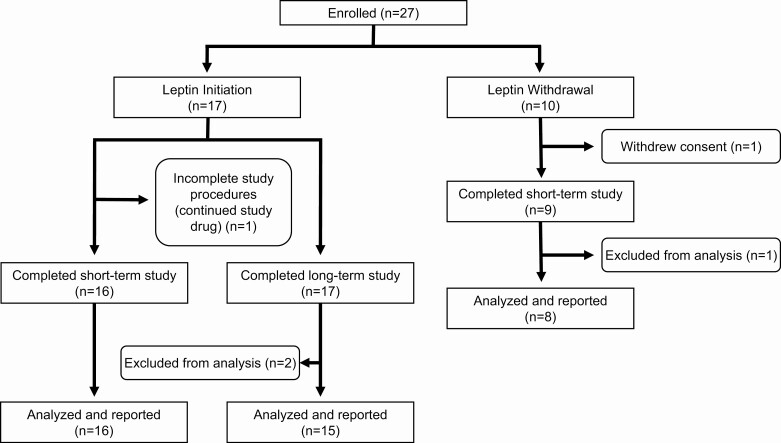
Design, setting, and patients: Nonrandomized crossover study of 25 patients with lipodystrophy (National Institutes of Health, 2013-2018).

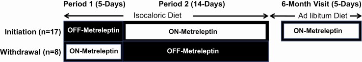
Intervention: The initiation cohort consisted of 17 patients without prior exposure to metreleptin, studied before and after 14 days of metreleptin. The withdrawal cohort consisted of 8 previously metreleptin-treated patients, studied before and after 14 days of metreleptin withdrawal.

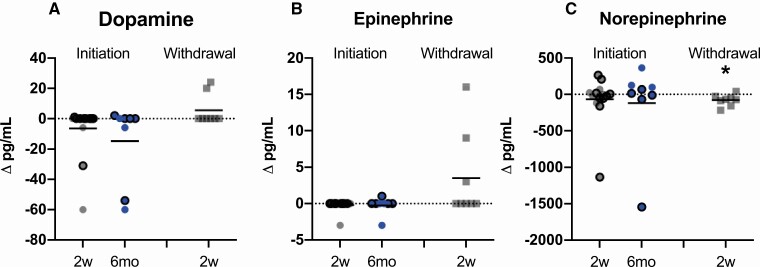
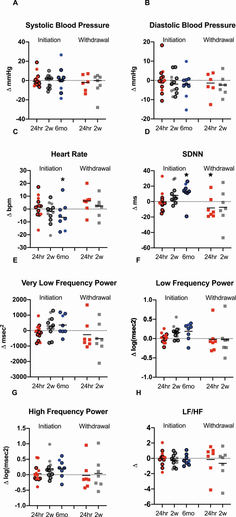
Main outcomes: 24-h total energy expenditure (TEE), resting energy expenditure (REE), autonomic nervous system activity [heart rate variability (HrV)], plasma-free triiodothyronine (T3), free thyroxine (T4), epinephrine, norepinephrine, and dopamine.

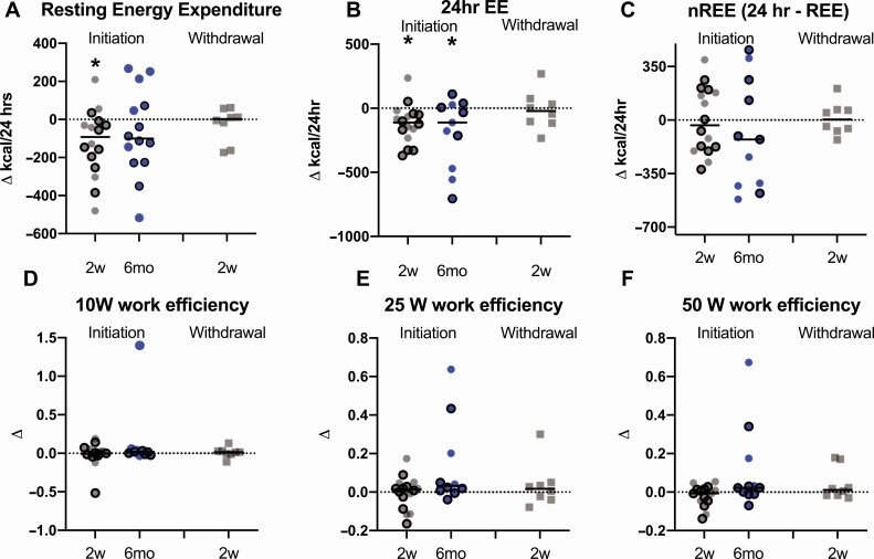
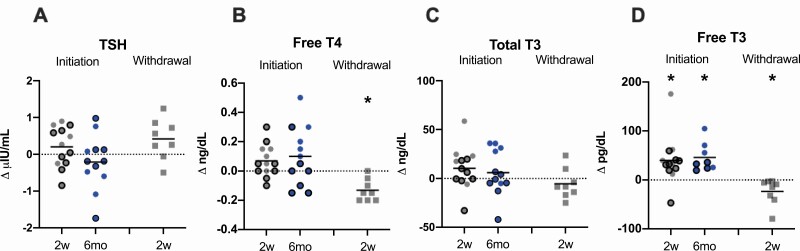
Results: In the initiation cohort, TEE and REE decreased by 5.0% (121 ± 152 kcal/day; P = 0.006) and 5.9% (120 ± 175 kcal/day; P = 0.02). Free T3 increased by 19.4% (40 ± 49 pg/dL; P = 0.01). No changes in catecholamines or HrV were observed. In the withdrawal cohort, free T3 decreased by 8.0% (P = 0.04), free T4 decreased by 11.9% (P = 0.002), and norepinephrine decreased by 34.2% (P = 0.03), but no changes in EE, epinephrine, dopamine, or HrV were observed.

Conclusions: Metreleptin initiation decreased EE in patients with lipodystrophy, but no changes were observed after metreleptin withdrawal. Thyroid hormone was higher on metreleptin in both initiation and withdrawal cohorts. Decreased EE after metreleptin in lipodystrophy may result from reductions in energy-requiring metabolic processes that counteract increases in EE via adipose tissue-specific neuroendocrine and adrenergic signaling.

Trial registration: ClinicalTrials.gov NCT01778556.

Keywords: autonomic nervous system; energy expenditure; leptin; lipodystrophy; thyroid hormone.


Figures

Figure 1.
Figure 1.
Study design schematic, nonrandomized, crossover study. Initiation patients (n = 17) were studied for the first 5 days without metreleptin (Period 1) and then treated with metreleptin for the next 14 days (Period 2). Withdrawal patients (n = 8) were studied for the first 5 days on their home dose of metreleptin (Period 1) and then withdrawn from metreleptin for the next 14 days (Period 2). Patients in the initiation cohort continued self-administered metreleptin treatment after discharge and were reevaluated after 6 months of metreleptin treatment on an ad libitum diet.
Figure 2.
Figure 2.
Study flow chart. Twenty-seven patients with generalized or partial lipodystrophy were enrolled in the study. Of the 17 patients who initiated metreleptin (initiation), 1 did not complete the study procedures for the short-term study (period 2) but was continued on metreleptin and re-evaluated at 6 months, and 1 completed the short-term study but was excluded from analysis of the 6-month data because of noncompliance with metreleptin. Eight patients who were taken off metreleptin for a 2-week period (withdrawal) completed the study and were analyzed.
Figure 3.
Figure 3.
Effects of metreleptin initiation or withdrawal on change in energy expenditure and skeletal muscle work efficiency in patients with lipodystrophy. Resting energy expenditure (A) decreased 2 weeks (2w; gray circles) after metreleptin initiation, but did not change from baseline to 6 months (6mo; blue circles) after metreleptin initiation or 2 weeks after metreleptin withdrawal (gray squares). Twenty-four-hour energy expenditure (B) decreased from baseline to 2 weeks and 6 months after metreleptin initiation but did not change after 2 weeks of metreleptin withdrawal. There were no changes in nonresting energy expenditure (C) or skeletal muscle work efficiency at 10 Watts (D), 25 Watts (E), and 50 Watts (F) from baseline to 2 weeks or 6 months following metreleptin initiation, or 2 weeks after metreleptin withdrawal (kcal/24 h). Patients with generalized lipodystrophy are shown as circles or squares without borders and those with partial lipodystrophy as circles with black borders. Comparisons were made using Wilcoxon rank-signed test and paired t-test for nonnormally and normally distributed data, respectively. Linear mixed effects models applying a Dunnett correction for multiple comparisons were used for outcomes measured at >2 time points. P < 0.05 represented statistical significance. P-values are 2-sided.
Figure 4.
Figure 4.
Effects of metreleptin initiation or withdrawal on catecholamines in patients with lipodystrophy. (A) Dopamine and (B) epinephrine did not change from baseline to 2 weeks (2w; gray circles) or 6 months (6mo; blue circles) after metreleptin initiation or 2 weeks after metreleptin withdrawal (2w; gray squares). (C) Norepinephrine did not change from baseline to 2 weeks or 6 months after metreleptin initiation but decreased 2 weeks following metreleptin withdrawal. Patients with generalized lipodystrophy are shown as circles or squares without borders and those with partial lipodystrophy as circles with black borders. Comparisons were made using Wilcoxon rank-signed test and paired t-test for nonnormally and normally distributed data, respectively. Linear mixed effects models applying a Dunnett correction for multiple comparisons were used for outcomes measured at >2 time points. P < 0.05 represented statistical significance. P-values are 2-sided.
Figure 5.
Figure 5.
Effects of metreleptin initiation or withdrawal on thyroid hormones in patients with lipodystrophy. (A) Thyroid stimulating hormone (TSH) did not change from baseline to 2 weeks (2w; gray circles) or 6 months (6mo; blue circles) after metreleptin administration or after 2 weeks (2w; gray squares) of metreleptin withdrawal. (B) Free thyroxine (T4) did not change from baseline to 2 weeks or 6 months after metreleptin administration but decreased after 2 weeks of metreleptin withdrawal. (C) Total triiodothyronine (T3) did not change at any time point from baseline following metreleptin initiation or withdrawal. (D) Free triiodothyronine (T3) increased from baseline to 2 weeks and 6 months after metreleptin initiation and decreased 2 weeks after metreleptin withdrawal. Patients with generalized lipodystrophy are shown as circles or squares without borders and those with partial lipodystrophy as circles with black borders. Comparisons were made using Wilcoxon rank-signed test and paired t-test for nonnormally and normally distributed data, respectively. Linear mixed effects models applying a Dunnett correction for multiple comparisons were used for outcomes measured at >2 time points. P < 0.05 represented statistical significance. P-values are 2-sided.
Figure 6.
Figure 6.
Effects of metreleptin initiation or withdrawal on blood pressure and heart rate variability metrics in patients with lipodystrophy. Systolic blood pressure (A) and diastolic blood pressure (B) did not change from baseline to 24 h (24hr; red circles), 2 weeks (2w; gray circles) or 6 months (6mo; blue circles) after metreleptin administration or after 24 h (24hr; red squares) or 2 weeks (2w; gray squares) following metreleptin withdrawal. Heart rate (C) did not change from baseline to 24 h or 2 weeks after metreleptin initiation but decreased at 6 months. No changes in heart rate were observed at any time point following metreleptin withdrawal. (D) SD of the normal-to-normal distance (SDNN) increased (associated with decreased cardiovascular disease risk) after 6 months of metreleptin initiation and decreased (associated with increased cardiovascular disease risk) after 24 h of metreleptin withdrawal. (E) The very low frequency (VLF) component of heart rate variability, a measure of renin-angiotensin-aldosterone system effects on heart rate variability, (F) low frequency power component (LF), a measure of both sympathetic and parasympathetic influence on heart rate variability, and (G) high frequency power component (HF), a measure of parasympathetic impact on heart rate variability, did not change from baseline to 24 h, 2 weeks, or 6 months after metreleptin initiation or after 24 h and 2 weeks following metreleptin withdrawal. (H) The LF/HF ratio, a measure of sympathetic influence on heart rate variability, did not change at any time point following metreleptin administration or withdrawal. Patients with generalized lipodystrophy are shown as circles or squares without borders and those with partial lipodystrophy as circles with black borders. Comparisons were made using Wilcoxon rank-signed test and paired t-test for normally and nonnormally distributed data. Linear mixed effects models applying a Dunnett correction for multiple comparisons were used for outcomes measured at >2 time points. P < 0.05 represented statistical significance. P-values are 2-sided.

References

    1. Friedman JM, Halaas JL. Leptin and the regulation of body weight in mammals. Nature. 1998;395(6704):763-770. - PubMed
    1. Pelleymounter MA, Cullen MJ, Baker MB, et al. . Effects of the obese gene product on body weight regulation in ob/ob mice. Science. 1995;269(5223):540-543. - PubMed
    1. Pandit R, Beerens S, Adan RAH. Role of leptin in energy expenditure: the hypothalamic perspective. Am J Physiol Regul Integr Comp Physiol. 2017;312(6):R938-R947. - PubMed
    1. Montague CT, Farooqi IS, Whitehead JP, et al. . Congenital leptin deficiency is associated with severe early-onset obesity in humans. Nature. 1997;387(6636):903-908. - PubMed
    1. Chan JL, Heist K, DePaoli AM, Veldhuis JD, Mantzoros CS. The role of falling leptin levels in the neuroendocrine and metabolic adaptation to short-term starvation in healthy men. J Clin Invest. 2003;111(9):1409-1421. - PMC - PubMed

Publication types

Associated data