. 2021 Jun 17;30(13):1230-1246.
doi: 10.1093/hmg/ddab116.

ALS/FTD mutations in UBQLN2 are linked to mitochondrial dysfunction through loss-of-function in mitochondrial protein import

Affiliations

ALS/FTD mutations in UBQLN2 are linked to mitochondrial dysfunction through loss-of-function in mitochondrial protein import

Brian C Lin et al. Hum Mol Genet. .

Abstract

UBQLN2 mutations cause amyotrophic lateral sclerosis (ALS) with frontotemporal dementia (FTD), but the pathogenic mechanisms by which they cause disease remain unclear. Proteomic profiling identified 'mitochondrial proteins' as comprising the largest category of protein changes in the spinal cord (SC) of the P497S UBQLN2 mouse model of ALS/FTD. Immunoblots confirmed P497S animals have global changes in proteins predictive of a severe decline in mitochondrial health, including oxidative phosphorylation (OXPHOS), mitochondrial protein import and network dynamics. Functional studies confirmed mitochondria purified from the SC of P497S animals have age-dependent decline in nearly all steps of OXPHOS. Mitochondria cristae deformities were evident in spinal motor neurons of aged P497S animals. Knockout (KO) of UBQLN2 in HeLa cells resulted in changes in mitochondrial proteins and OXPHOS activity similar to those seen in the SC. KO of UBQLN2 also compromised targeting and processing of the mitochondrial import factor, TIMM44, resulting in accumulation in abnormal foci. The functional OXPHOS deficits and TIMM44-targeting defects were rescued by reexpression of WT UBQLN2 but not by ALS/FTD mutant UBQLN2 proteins. In vitro binding assays revealed ALS/FTD mutant UBQLN2 proteins bind weaker with TIMM44 than WT UBQLN2 protein, suggesting that the loss of UBQLN2 binding may underlie the import and/or delivery defect of TIMM44 to mitochondria. Our studies indicate a potential key pathogenic disturbance in mitochondrial health caused by UBQLN2 mutations.


Figures

Figure 1
Figure 1
Mitochondrial protein changes in the P497S UBQLN2 mouse model of ALS/FTD. (A) Immunoblots of SC lysates from three independent mice for the three genotypes probed for mitochondrial electron transport chain proteins. (B) Quantification of the blots shown in A. *P < 0.05, **P < 0.01. (C) Immunoblots of SC lysates from 32-week-old animals probed for mitochondrial dynamics and import proteins. (D) Quantification of the blots shown in C. *P < 0.05, **P < 0.01, ***P < 0.001. (E) Immunoblots of SC lysates from mice of different ages and genotypes probed for Opa1. (F) Quantification of uncleaved Opa1 (100 kDa) relative to total Opa1 levels. *P < 0.05, ***P < 0.001.
Figure 2
Figure 2
Structural and functional mitochondrial defects in the SC of P497S animals. (A) Immunoblots of equal protein lysates of mitochondria purified from the SC from four 52-week-old animals for the three genotypes probed for the proteins shown. (B) Quantification of the blots shown in A. *P < 0.05, **P < 0.01, ***P < 0.001. (C) Seahorse mitochondria respiration assays of freshly isolated mitochondria from the lumbar SC of mice from the three genotypes at 24, 32 and 52 weeks of age; oxygen consumption rate (OCR) curves are shown. n = 3–5. (D) Bar charts depict basal, State 3 (ADP-stimulated), State 4o (proton leak) and State 3u (maximal) respiration rates from traces shown in C. *P < 0.05, **P < 0.01, ***P < 0.001. (E) TEM images of the cell body regions of MN in the SC of 52-week-old mice for the three genotypes. Also shown are the magnified images of particular mitochondria in the MN (arrows). Scale bars for representative images, 500 and 100 nm for zoomed in mitochondria. (F) Average mitochondria length (μm) in SC motor neurons. **P < 0.01, ****P < 0.0001.
Figure 3
Figure 3
Inactivation of UBQLN2 expression in cells recapitulates the mitochondrial deficits seen in P497S animals. (A) Immunoblots of four independent lysates from parental HeLa WT and CRISPR/cas9 UBQLN2 KO8 lines probed for the indicated mitochondrial import and OXPHOS proteins. (B) Quantification of the blots shown in A. **P < 0.01, ***P < 0.001. (C) OCR curves depicting respiration rates of HeLa WT and UBQLN2 KO8 cell lines grown in standard DMEM media supplemented with glucose. (D) Bar chart of basal, ATP (oligomycin-sensitive) and maximal respiration. n = 4, ns, not significant. (E) OCR curves of HeLa WT and UBQLN2 KO8 lines grown in oxidative DMEM media supplemented with galactose. (F) Bar chart of basal, ATP (oligomycin-sensitive) and maximal respiration. n = 4, ***P < 0.001.
Figure 4
Figure 4
Alterations in mitochondria proteins, structure, function and dynamics in UBQLN2 KO cells and rescue of the functional deficits by reexpression of WT UBQLN2, but not by mutant P497S UBQLN2 proteins. (A) Parental HeLa WT and UBQLN2 KO8 lines stained with Tom20 to decorate mitochondria. Scale bar, 10 μm. (B) Quantification of the mitochondria network morphology of WT and KO8 cells into the percentage with tubular (>80% elongated mitochondria), intermediate (>50% short rod-like mitochondria) or fragmented (>50% fragmented mitochondria) mitochondria. n = 5, 50 cells per group. (C) Immunoblots of four independent lysates from WT and KO8 cultures probed for proteins shown. (D) Quantification of mitochondrial dynamic proteins shown in C. *P < 0.05, **P < 0.01, ***P < 0.001. (E) Time course images showing the extent of dissipation of GFP fluorescence in two focally activated regions in WT and KO8 cells expressing the mito-PAGFP reporter. (F) Quantification of the time of first fusion from the assay shown in E. n = 12, **P < 0.01. (G) OCR curves depicting parental HeLa WT and UBQLN2 KO8 cultures transfected with empty vector control, HA-tagged WT UBQLN2 or HA-tagged P497S UBQLN2 grown in oxidative DMEM media supplemented with galactose. (H) Quantification of basal and maximal respiration are shown for traces in G. n = 5, *P < 0.05, **P < 0.01, ***P < 0.001. (I) Immunoblots to show expression of the HA-tagged UBQLN2 constructs for the three separate experiments shown in G and H. (J) Quantification of HA-tagged WT and P497S mutant UBQLN2 proteins shown in I.
Figure 5
Figure 5
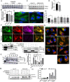
Alteration of TIMM44 protein levels in P497S animals and differences in mitochondrial targeting of the protein in cells containing and lacking UBQLN2. (A) Immunoblots of SC lysates from three independent 32-week-old animals for the three mouse genotypes probed for TIMM44 and GAPDH. (B) Quantification of TIMM44 levels in the blots shown in A after normalization of protein loading. *P < 0.05. (C) Immunoblot of four different HeLa WT and UBQLN2 KO8 cultures probed for TIMM44 and GAPDH. (D) Same as B, but for the blots shown in C. *P < 0.05. (E) Representative images of TIMM44-GFP fluorescence and of DAPI staining in HeLa WT and UBQLN2 KO8 cells. Scale bar, 10 μm. (F) Different color fluorescence images of cells stained for the proteins shown to monitor targeting of TIMM44-GFP to mitochondria in HeLa WT (1st row) and UBQLN2 KO8 cells (2nd row). Magnified images of boxed area in the 1st row are shown on the upper right-hand side. Scale bar, 10 and 5 μm. (G) Merged images of KO8 cells showing extent of colocalization of GFP fluorescence of the TIMM44-GFP reporter with TOMM20 staining in cells cotransfected with empty vector, or with WT UBQLN2, or with five different ALS/FTD mutant UBQLN2 cDNAS. Scale bar, 10 μm. (H) Quantification of cells with TIMM44 foci for experiment presented in G. n = 150–300 cells, *P < 0.05. (I) Immunoprecipitation of endogenous proteins from HeLa cells with either IgG control antibody or anti-UBQLN2 antibody and immunoblotted for either TIMM44 (short and long exposures) or UBQLN2. (J) Pulldown assays showing direct binding between both FL and the ΔLS TIMM44-HIS recombinant proteins with GST-UBQLN2 fusion protein. (K) Same as J, but comparing binding of FL TIMM44-HIS protein with WT and ALS/FTD mutant GST-fusion proteins. (L) Quantification of the amount of TIMM44 pulldown relative to the amount of GST-fusion protein recovered and normalized to the binding of WT GST-UBQLN2 protein. n = 4, **P < 0.01. ***P < 0.001. (M) Demonstration that UBQLN2 promotes maturation of TIMM44. HeLa and UBQLN2 KO8 cultures were transfected with cDNAs (μg) as shown (equivalent amount of total cDNA). Lysates were immunoblotted and probed with anti-Myc and anti-HA antibodies. (N) Quantification of precursor TIMM44 (50 kDa) over total TIMM44 levels in blot presented in M.

References

    1. Deng, H.X., Chen, W., Hong, S.T., Boycott, K.M., Gorrie, G.H., Siddique, N., Yang, Y., Fecto, F., Shi, Y., Zhai, H. et al. (2011) Mutations in UBQLN2 cause dominant X-linked juvenile and adult-onset ALS and ALS/dementia. Nature, 477, 211–215. - PMC - PubMed
    1. Higgins, N., Lin, B. and Monteiro, M.J. (2019) Lou Gehrig's disease (ALS): UBQLN2 mutations strike out of phase. Structure, 27, 879–881. - PMC - PubMed
    1. Mah, A.L., Perry, G., Smith, M.A. and Monteiro, M.J. (2000) Identification of ubiquilin, a novel presenilin interactor that increases presenilin protein accumulation. J. Cell Biol., 151, 847–862. - PMC - PubMed
    1. Marin, I. (2014) The ubiquilin gene family: evolutionary patterns and functional insights. BMC Evol. Biol., 14, 63. - PMC - PubMed
    1. Rothenberg, C. and Monteiro, M.J. (2010) Ubiquilin at a crossroads in protein degradation pathways. Autophagy, 6, 979–980. - PMC - PubMed

Publication types

MeSH terms

Substances