Review
. 2022 Feb;45(1):1-25.
doi: 10.1007/s10143-021-01543-z. Epub 2021 Apr 23.

The evolution of intracranial aneurysm treatment techniques and future directions

Affiliations
Review

The evolution of intracranial aneurysm treatment techniques and future directions

Keng Siang Lee et al. Neurosurg Rev. 2022 Feb.

Abstract

Treatment techniques and management guidelines for intracranial aneurysms (IAs) have been continually developing and this rapid development has altered treatment decision-making for clinicians. IAs are treated in one of two ways: surgical treatments such as microsurgical clipping with or without bypass techniques, and endovascular methods such as coiling, balloon- or stent-assisted coiling, or intravascular flow diversion and intrasaccular flow disruption. In certain cases, a single approach may be inadequate in completely resolving the IA and successful treatment requires a combination of microsurgical and endovascular techniques, such as in complex aneurysms. The treatment option should be considered based on factors such as age; past medical history; comorbidities; patient preference; aneurysm characteristics such as location, morphology, and size; and finally the operator's experience. The purpose of this review is to provide practicing neurosurgeons with a summary of the techniques available, and to aid decision-making by highlighting ideal or less ideal cases for a given technique. Next, we illustrate the evolution of techniques to overcome the shortfalls of preceding techniques. At the outset, we emphasize that this decision-making process is dynamic and will be directed by current best scientific evidence, and future technological advances.

Keywords: Aneurysms; Clipping; Coiling; Endovascular embolization; Flow diversion; Stents; Subarachnoid hemorrhage.


Conflict of interest statement

The authors declare no competing interests.

Figures

Fig. 1
Fig. 1
A large right ruptured MCA aneurysm was treated with a total of 19 coils. The red arrow points to an incidental right paraclinoid unruptured aneurysm
Fig. 2
Fig. 2
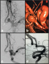
Ruptured, wide-neck 4-mm right PCommA aneurysm in a poor surgical candidate. a AP and b lateral views. The initial framing coil would not stay in the aneurysm; thus, a balloon was inflated during placement of the initial framing coil allowing it to c remain within the aneurysm. d AP and e lateral views after subsequent coils were placed obliterating the dome. Dome remains obliterated at 2-year follow-up
Fig. 3
Fig. 3
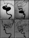
Unruptured 7-mm, wide-necked ACommA aneurysm. a AP view demonstrating Y configuration stents with distal markers visible in each A2 segment (arrows) and proximal maker in the A1 (arrow) and b a 3D view of the neck. Post-coiling c AP and d lateral view demonstrating aneurysm obliteration without encroachment of the parent vessels
Fig. 4
Fig. 4
a AP and b lateral pre-treatment angiogram demonstrating a giant left cavernous segment ICA aneurysm. c AP and d lateral projections of the left ICA 6 months after flow diversion treatment demonstrating resolution of the aneurysm and remodeling of the parent vessel
Fig. 5
Fig. 5
Pre-treatment a AP, b lateral, and c 3D images of a right anterior temporal artery. MCA aneurysm with the anterior temporal artery originating from the neck (arrow). Immediate post-WEB device deployment d unsubtracted image showing the device and e subtracted lateral and image demonstrating aneurysm obliteration with patency of the branching artery (arrow). Six-month follow-up f AP image showing continued occlusion with patent anterior temporal artery (arrow)
Fig. 6
Fig. 6
Clipping of a partially calcified MCA bifurcation aneurysm is demonstrated here with temporary clipping of the M1 trunk to soften the aneurysm dome. In the upper left corner, a permanent clip is seen on a posterior communicating artery (PCommA) aneurysm (not shown) that was concomitantly clipped during the same operation
Fig. 7
Fig. 7
An 18-year-old male presented with intermitted left-sided weakness. a MRI brain showed ischemic changes within the right MCA territory with wide-necked, complex MCA aneurysm. b Preoperative cerebral angiogram (right ICA injection) showed complex MCA aneurysm with MCA branches arising from the body of the aneurysm. c Right ECA-saphenous vein-MCA bypass (EC-IC bypass) was performed, with postoperative cerebral angiogram confirmed right MCA perfusion by high-flow bypass graft. d Schematic diagram to illustrate the complex aneurysm configuration, and bypass strategy, implanting the MCA branches into the saphenous vein graft. e, f Intraoperative view of the cranial incision, neck incision, and right saphenous vein harvest. He made good postoperative recovery, and resumed surfing
Fig. 8
Fig. 8
A 65-year-old man, hypertensive, smoker, and with previous TIAs, was diagnosed with a large right MCA aneurysm 13 years prior that was managed conservatively (a). He presented with progressive left hemiparesis, impaired conscious level. b CT head showed a heavily calcified giant right MCA aneurysm with surrounding edema. c, d CT angiogram (axial and coronal views) showed patent aneurysm remnant, heavy calcification at the aneurysm neck, and thick layers of intraaneurysmal thrombus. e MRI brain, T2-weighted, axial view showed onion-ring appearance of multilayered intraaneurysmal thrombus. f 3D reconstructed angiogram showed single MCA branch arising from the aneurysm neck. g He underwent aneurysmectomy with in situ MCA end-to-end anastomosis using interposition saphenous vein graft (IC-IC bypass) and made good recovery. At 6 months follow-up, he was independently mobile, with resolution of hemiparesis. g, h CT angiogram confirmed patent in situ bypass graft (red arrows)
Fig. 9
Fig. 9
A 10-year-old patient presented with coma producing aneurysmal SAH. Preintubation, his best motor score was flexing. a CT head showed diffused SAH, with associated frontal hematoma. b CT angiogram confirmed an underlying complex multilobulated anterior communicating artery (ACommA) aneurysm. c Cerebral angiogram (R ICA injection) showed the multilobulated ACommA, and right A2 arising from the neck of the aneurysm. He underwent coil occlusion of the ruptured ACommA, securing the ruptured fundal component. d MR angiogram, small neck remnant of coiled aneurysm was evident to protect the right A2 branch. After a period of ICU stay and rehabilitation, he made good recovery. e MR angiogram at 6 months follow-up showed significant aneurysm recurrence. f Cerebral angiogram (left ICA) injection showed ACommA aneurysm enlargement. After multidisciplinary discussion, we proceeded to perform right pericallosal to left pericallosal in situ (IC-IC) bypass, prior to further coil occlusion of the aneurysm, sacrificing the right A2. g Intraoperative view of pericallosal-pericallosal in situ bypass (IC-IC), end to side technique using interrupted 9/0 suture. h Angiogram after further coil occlusion of the recurrent ACommA aneurysm, and left A2 supplying both pericallosal branches (red arrow)
Fig. 10
Fig. 10
A 50-year-old patient with poor-grade SAH, treated by hybrid approach. a Cerebral angiogram (right ICA injection) confirmed a complex right MCA aneurysm. b, c Post-coiling angiogram showed satisfactory aneurysm occlusion, with protection of the fundal bleeding point. d Six months follow-up angiogram showed aneurysm neck recurrence/coil compaction. e, f 3-Dimensional reconstructed angiogram (AP, PA views) showed the complex wide-necked MCA aneurysm configuration with multiple daughter sacs. g Post-clipping angiogram confirmed complete right MCA aneurysm obliteration, using h clip reconstruction technique preserving MCA branches (i), via mini-pterional approach

References

    1. Aboukais R, Zairi F, Bourgeois P, Boustia F, Leclerc X, Lejeune JP. Pericallosal aneurysm: a difficult challenge for microsurgery and endovascular treatment. Neurochirurgie. 2015;61:244–249. - PubMed
    1. Albuquerque FC, Gonzalez LF, Hu YC, Newman CB, McDougall CG. Transcirculation endovascular treatment of complex cerebral aneurysms: technical considerations and preliminary results. Neurosurgery. 2011;68:820–829. - PubMed
    1. Alreshidi M, Cote DJ, Dasenbrock HH, Acosta M, Can A, Doucette J, Simjian T, Hulou MM, Wheeler LA, Huang K, Zaidi HA, Du R, Aziz-Sultan MA, Mekary RA, Smith TR. Coiling versus microsurgical clipping in the treatment of unruptured middle cerebral artery aneurysms: a meta-analysis. Neurosurgery. 2018;83:879–889. - PubMed
    1. AngioCalc, AngioCalc Cerebral and Peripheral Aneurysm Calculator.
    1. Arthur AS, Molyneux A, Coon AL, Saatci I, Szikora I, Baltacioglu F, Sultan A, Hoit D, Delgado Almandoz JE, Elijovich L, Cekirge S, Byrne JV, Fiorella D, W.-I.S. investigators The safety and effectiveness of the Woven EndoBridge (WEB) system for the treatment of wide-necked bifurcation aneurysms: final 12-month results of the pivotal WEB Intrasaccular Therapy (WEB-IT) Study. J Neurointerv Surg. 2019;11:924–930. - PMC - PubMed