. 2021 Jul;43(7):1467-1473.
doi: 10.1007/s10529-021-03135-9. Epub 2021 Apr 23.

A single point mutation converts a glutaryl-7-aminocephalosporanic acid acylase into an N-acyl-homoserine lactone acylase

Affiliations

A single point mutation converts a glutaryl-7-aminocephalosporanic acid acylase into an N-acyl-homoserine lactone acylase

Shereen A Murugayah et al. Biotechnol Lett. 2021 Jul.

Abstract

Objective: To change the specificity of a glutaryl-7-aminocephalosporanic acid acylase (GCA) towards N-acyl homoserine lactones (AHLs; quorum sensing signalling molecules) by site-directed mutagenesis.

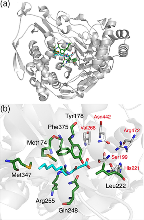
Results: Seven residues were identified by analysis of existing crystal structures as potential determinants of substrate specificity. Site-saturation mutagenesis libraries were created for each of the seven selected positions. High-throughput activity screening of each library identified two variants-Arg255Ala, Arg255Gly-with new activities towards N-acyl homoserine lactone substrates. Structural modelling of the Arg255Gly mutation suggests that the smaller side-chain of glycine (as compared to arginine in the wild-type enzyme) avoids a key clash with the acyl group of the N-acyl homoserine lactone substrate.

Conclusions: Mutation of a single amino acid residue successfully converted a GCA (with no detectable activity against AHLs) into an AHL acylase. This approach may be useful for further engineering of 'quorum quenching' enzymes.

Keywords: Glutaryl-7-aminocephalosporanic acid acylase; N-acyl-homoserine lactone acylase; Protein engineering; Quorum quenching; Site‐saturation mutagenesis.


Conflict of interest statement

The authors declare they have no conflict of interest.

Figures

Fig. 1
Fig. 1
Selection of GCA residues for mutagenesis a Ribbon diagram of the overall structural fold of GCA (grey) with residues selected for mutagenesis shown as green sticks. 3-oxododecanoic acid (3-oxo-C12; cyan sticks) was modelled into the GCA structure by aligning it with the 3-oxo-C12 bound-PvdQ structure. b The GCA active site with 3-oxo-C12 (cyan sticks). Non-catalytic residues within a 5 Å radius of the modelled substrate were chosen for mutagenesis; these are shown as sticks with green backbones. Residues that are critical for catalysis are shown as sticks with white backbones. PDB entries for GCA [1OR0 (Kim et al. 2000)] and PvdQ [PDB 2WYC (Bokhove et al. 2010)] were used to construct the figure using PyMOL (Schrödinger, LLC)
Fig. 2
Fig. 2
Modelling of the active sites of with GCA and the Arg255Gly variant. a Wild-type GCA with its native GL7-ACA substrate. b Wild-type GCA with 3-oxo-C12-HSL. c Arg255Gly GL7-ACA. d Arg255Gly with 3-oxo-C12. Residue 255 is shown as green sticks. Substrates (GL7-ACA and 3-oxo-C12-HSL) are shown as pink sticks. Residues that are critical for catalysis are shown as sticks with white backbones
Fig. 3
Fig. 3
The effect of enzyme treatment on P. aeruginosa biofilm formation. Biofilms of P. aeruginosa PAO1 (Pa PAO1, dark grey) and P. aeruginosa clinical isolate (Pa CI, light grey) were grown in the presence of the wild-type GCA (wild-type) or Arg255Gly mutant (Arg255Gly) and biofilm formation was measured using crystal violet staining of the bacterial biomass. Two biological replicates were performed (n = 2) with four technical replicates each. The independent data points are shown as circles, bars represent the mean, and lines represent the range

References

    1. Billot R, Plener L, Jacquet P, Elias M, Chabriere E, Daude D. Engineering acyl-homoserine lactone-interfering enzymes toward bacterial control. J Biol Chem. 2020;295:12993–13007. doi: 10.1074/jbc.REV120.013531. - DOI - PMC - PubMed
    1. Bokhove M, Jimenez PN, Quax WJ, Dijkstra BW. The quorum-quenching N-acyl homoserine lactone acylase PvdQ is an Ntn-hydrolase with an unusual substrate-binding pocket. Proc Natl Acad Sci USA. 2010;107:686–691. doi: 10.1073/pnas.0911839107. - DOI - PMC - PubMed
    1. Conti G, Pollegioni L, Molla G, Rosini E. Strategic manipulation of an industrial biocatalyst–evolution of a cephalosporin C acylase. FEBS J. 2014;281:2443–2455. doi: 10.1111/febs.12798. - DOI - PubMed
    1. Fetzner S. Quorum quenching enzymes. J Biotechnol. 2015;201:2–14. doi: 10.1016/j.jbiotec.2014.09.001. - DOI - PubMed
    1. Firth AE, Patrick WM. GLUE-IT and PEDEL-AA: new programmes for analyzing protein diversity in randomized libraries. Nucleic Acids Res. 2008;36:W281–W285. doi: 10.1093/nar/gkn226. - DOI - PMC - PubMed

MeSH terms

LinkOut - more resources