. 2021 Sep 14;16(9):2213-2227.
doi: 10.1016/j.stemcr.2021.03.029. Epub 2021 Apr 22.

Human motor units in microfluidic devices are impaired by FUS mutations and improved by HDAC6 inhibition

Affiliations

Human motor units in microfluidic devices are impaired by FUS mutations and improved by HDAC6 inhibition

Katarina Stoklund Dittlau et al. Stem Cell Reports. .

Abstract

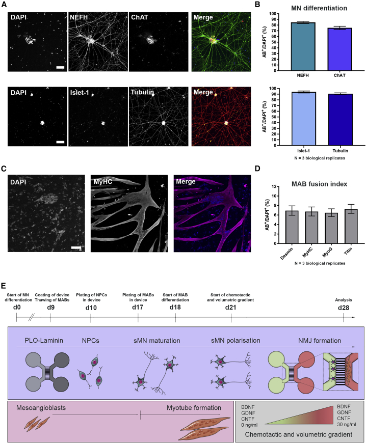
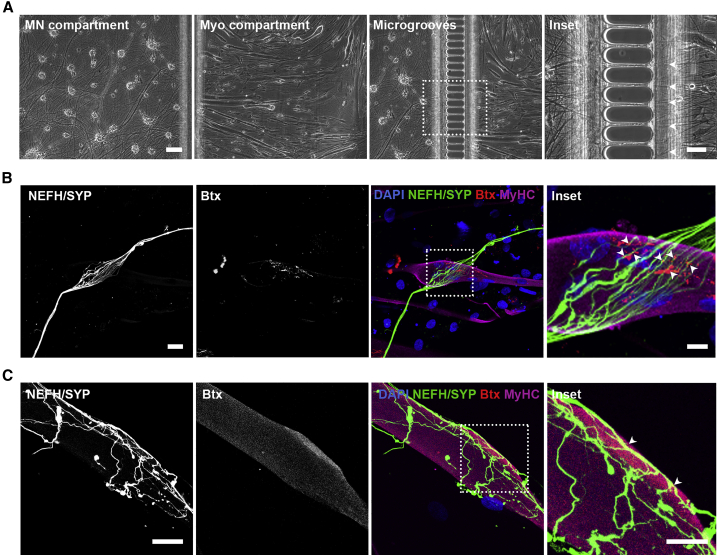
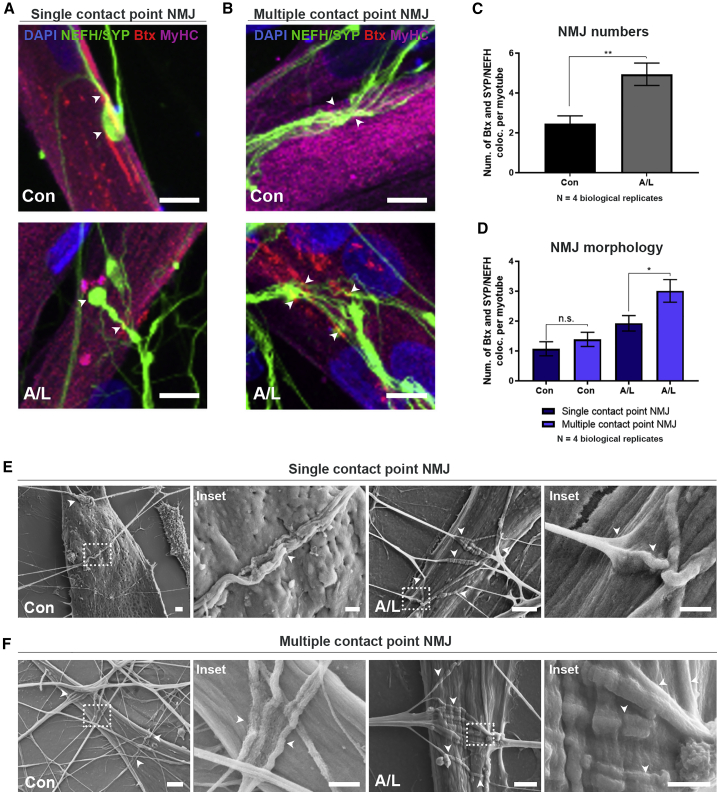
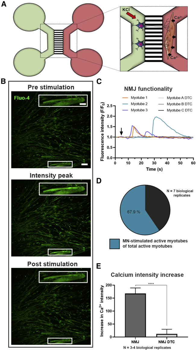
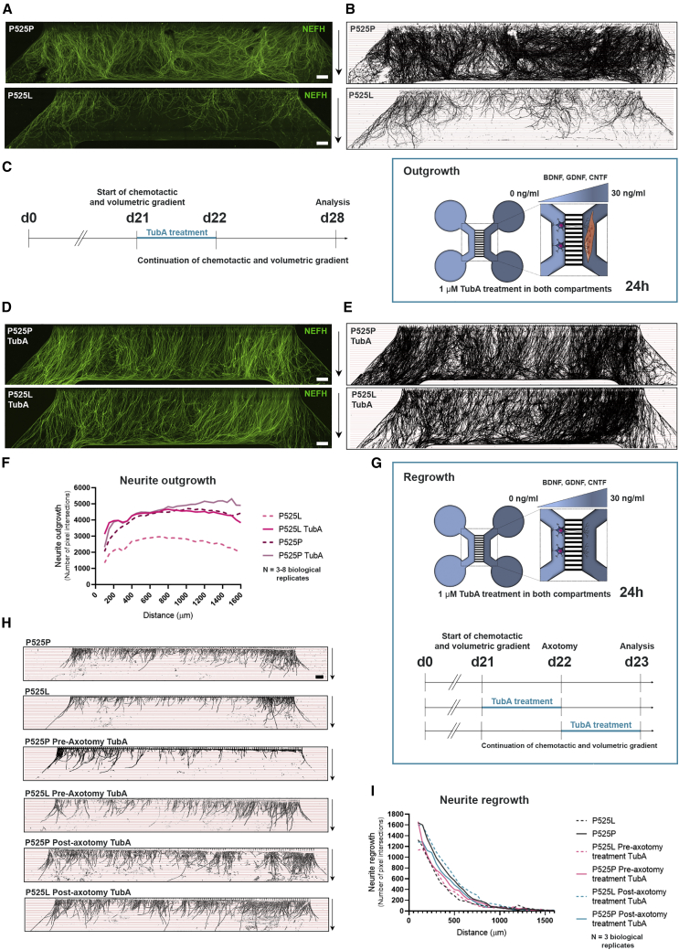
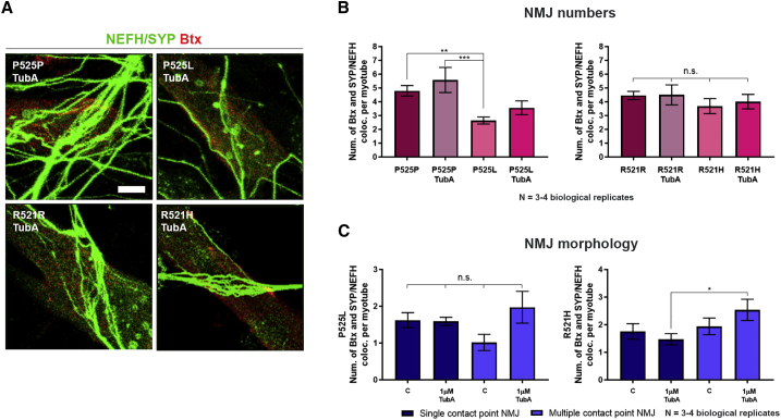
Neuromuscular junctions (NMJs) ensure communication between motor neurons (MNs) and muscle; however, in MN disorders, such as amyotrophic lateral sclerosis (ALS), NMJs degenerate resulting in muscle atrophy. The aim of this study was to establish a versatile and reproducible in vitro model of a human motor unit to investigate the effects of ALS-causing mutations. Therefore, we generated a co-culture of human induced pluripotent stem cell (iPSC)-derived MNs and human primary mesoangioblast-derived myotubes in microfluidic devices. A chemotactic and volumetric gradient facilitated the growth of MN neurites through microgrooves resulting in the interaction with myotubes and the formation of NMJs. We observed that ALS-causing FUS mutations resulted in reduced neurite outgrowth as well as an impaired neurite regrowth upon axotomy. NMJ numbers were likewise reduced in the FUS-ALS model. Interestingly, the selective HDAC6 inhibitor, Tubastatin A, improved the neurite outgrowth, regrowth, and NMJ morphology, prompting HDAC6 inhibition as a potential therapeutic strategy for ALS.

Keywords: FUS; HDAC6; Tubastatin A; amyotrophic lateral sclerosis; microfluidic device; neurite outgrowth; neurite regrowth; neuromuscular junction.


Conflict of interest statement

L.V.D.B. has a patent on the use of HDAC inhibitors in Charcot-Marie-Tooth disease (US-2013227717-A1), is scientific co-founder of Augustine Therapeutics and a member of its scientific advisory board. The other authors declare no competing interests.

Figures

Figure 1
Figure 1
Characterization of monocultures and overview of NMJ protocol (A) Confocal images of MNs stained with MN markers neurofilament heavy chain (NEFH), choline acetyltransferase (ChAT), and Islet-1, as well as the pan-neuronal marker βIII-tubulin (Tubulin) at day 28 of MN differentiation. Nuclei stained with DAPI. Scale bars, 75 μm. (B) Number of cells positive for MN and pan-neuronal markers (AB+). Mean ± SEM of three biological replicates. (C) Confocal images of myotube heavy chain (MyHC)-positive myotubes 10 days after initiation of differentiation. Scale bar, 75 μm. (D) Quantification of mesoangioblast (MAB) fusion into multinucleated myotubes (fusion index) with myotube markers desmin, MyHC, myogenin (MyoG) or titin. Mean ± SEM of three biological replicates. (E) Schematic overview of co-culture protocol and differentiation timeline (days 0–28). Day 0 (d0), differentiation of iPSCs into MN. Day 9 (d9), microfluidic devices are coated with poly-L-ornithine (PLO) and laminin, and MABs are thawed for expansion. Day 10 (d10), MN-NPCs are plated on one side (light gray) of the device. Day 17 (d17), MABs are seeded in the opposite side of the device (dark gray). Myotube differentiation is initiated (d18). Day 21 (d21), a volumetric and chemotactic gradient of neurotrophic factors (BDNF, GDNF and CNTF) is implemented to facilitate polarized growth of spinal MNs (sMN) through the microgrooves toward the myotubes to initiate the formation of NMJs (d28). Cell illustrations were modified from Smart Servier Medical Art licensed under a Creative Commons Attribution 3.0 Unported License (https://creativecommons.org/licenses/by/3.0/). See also Figure S1.
Figure 2
Figure 2
Co-culturing of MNs and myotubes in microfluidic devices leads to NMJ formation (A) Bright-field micrographs of MN and myotube (Myo) compartments in the XC150 device on day 28. Insets: magnification of axonal migration through microgrooves (arrowheads). Scale bars, 100 μm and 50 μm (insets). (B and C) Confocal micrograph of NMJ formation are shown as co-localization of presynaptic marker synaptophysin (SYP) and acetylcholine receptor marker α-bungarotoxin (Btx) on MyHC-labeled myotubes. Insets: magnification of co-localizations (arrowheads). Scale bars, 25 μm and 10 μm (insets).
Figure 3
Figure 3
Agrin and laminin improve NMJ formation in microfluidic devices (A and B) Confocal micrographs of NMJs in agrin (0.01 μg/mL)- and laminin (20 μg/mL)-supplemented (A/L) and untreated (Con) conditions on day 28 in XC150 devices. Single contact point NMJs (A). Multiple contact point NMJs (B). Scale bars, 10 μm. Arrowheads: Btx-SYP/NEFH co-localizations. (C) Number of Btx and SYP/NEFH co-localizations per myotube. (D) Quantifications of NMJ single and multiple contact point morphology. (E and F) Representative scanning electron microscopy (SEM) images of NMJ formation in SND75 devices on day 28 in A/L-supplemented and control conditions. Single contact point NMJs (E). Multiple contact point NMJs (F). Insets: magnifications of NMJ (arrowheads). Scale bars, 2 μm and 1 μm (insets). (C) Unpaired t test. (D) One-way ANOVA with Tukey's multiple comparisons test. Mean ± SEM of four biological replicates. p < 0.05, ∗∗p < 0.01. See also Figures S2 and S3.
Figure 4
Figure 4
In vitro NMJs are functionally active (A) Schematic overview of transient fluorescent Ca2+ imaging in XC150 devices on day 28. MN compartment (green) is stimulated with 50 mM KCl, which initiates an intracellular response through MN axons evoking an increase in Ca2+ influx in the Fluo-4-loaded myotubes (red compartment). Cell illustrations are modified from Smart Servier Medical Art licensed under a Creative Commons Attribution 3.0 Unported License (https://creativecommons.org/licenses/by/3.0/). (B) Fluorescent micrograph examples of before, during, and after stimulation depicting a wave of increase in intracellular Ca2+ in myotubes upon MN stimulation. Inset shows a magnification of an innervated active myotube. Scale bars, 100 μm and 200 μm (insets). (C) Representative Ca2+ influx curves in myotubes after KCl activation (arrow) of MNs before (myotube 1–3) and after 10 min treatment with NMJ blocker tubocurarine (myotube A-C DTC). (D) Ratio of MN-stimulated active myotubes to directly KCl-activated myotubes. (E) Effect of DTC on the intensity fluorescent increase due to Ca2+ influx induced by KCl increase in the MN compartment. Mean ± SEM of three to four biological replicates. Mann-Whitney test. ∗∗∗∗p < 0.0001. See also Figure S4.
Figure 5
Figure 5
NMJs are impaired in ALS (A) Confocal micrographs of NMJs with A/L from ALS-FUS (P525L, R521H) and isogenic control (P525P, R521R) MN/myotube co-cultures on day 28. Scale bars, 10 μm. Arrowheads: Btx-SYP/NEFH co-localizations. (B) Quantification of NMJs as Btx and SYP/NEFH co-localizations and expressed per myotube. (C) Quantification of NMJ single and multiple contact point morphology. Mean ± SEM of three to four biological replicates. (B) Unpaired t test or Mann-Whitney test. (C) Kruskal-Wallis test with Dunn's multiple comparisons test. p < 0.05, ∗∗∗p < 0.001, ∗∗∗∗p < 0.0001. See also Figures S4, S5, and S7.
Figure 6
Figure 6
ALS-dependent impairment in neurite outgrowth and regrowth is rescued by HDAC6 inhibition (A) Tile scan confocal overviews of neurite outgrowth (NEFH) in the myotube compartment from P525L and P525P cultures at day 28. Arrows (right): growth direction from exit of microgrooves. Scale bars, 300 μm. (B) Masks of tile scans with intersection lines at every 50 μm starting from microgroove exit. (C) Schematic representation of outgrowth experimental time course implementing treatment with TubA. At day 21 (d21), both MN and myotube compartments were treated with 1 μM TubA for 24 h in addition to the start of the chemotactic and volumetric gradient. (D) Tile scan confocal overviews of neurite outgrowth in myotube compartment at day 28 from P525L and P525P MN/myotube co-cultures with TubA treatment. Scale bars, 300 μm. (E) Masks of tile scans with TubA treatment. (F) Neurite outgrowth quantifications of the number of pixel intersections in P525L and P525P MN/myotube co-cultures with and without TubA treatment. Mean graph of three to eight biological replicates. (G) Schematic representation of regrowth experimental time course implementing 24 h treatment with TubA before or after axotomy (day 22). (H) Masks of tile scan overviews after 24 h neurite regrowth in empty myotube compartment from P525L and P525P cultures without, and with pre-axotomy and post-axotomy treatment with TubA. Arrows (right): growth direction from exit of microgrooves. Scale bar, 300 μm. (I) Neurite regrowth quantifications of the number of pixel intersections. Mean graph of three biological replicates. Cell illustrations in (C and G) are modified from Smart Servier Medical Art licensed under a Creative Commons Attribution 3.0 Unported License (https://creativecommons.org/licenses/by/3.0/). See also Figures S4, S6, and S7 and Tables S3 and S4.
Figure 7
Figure 7
HDAC6 inhibition improves ALS-dependent impairments in NMJ morphology (A) Confocal micrographs of NMJs with TubA treatment. Scale bar, 10 μm. (B) Quantification of NMJs as Btx-SYP/NEFH co-localization per myotube with and without TubA treatment. Control data without TubA treatment are identical to Figure 5B. (C) Morphological analysis of NMJs without (C) and with TubA treatment. Control data (C) are identical to Figure 5C. Mean ± SEM from three to four biological replicates. One-way ANOVA with Tukey's multiple comparisons test or Kruskal-Wallis test with Dunn's multiple comparisons test. p < 0.05, ∗∗p < 0.01, ∗∗∗p < 0.001. See also Figures S4 and S7.

References

    1. Afshar Bakooshli M., Lippmann E.S., Mulcahy B., Iyer N., Nguyen C.T., Tung K., Stewart B.A., van den Dorpel H., Fuehrmann T., Shoichet M., et al. A 3D culture model of innervated human skeletal muscle enables studies of the adult neuromuscular junction. eLife. 2019;8:e44530. - PMC - PubMed
    1. Akiyama T., Suzuki N., Ishikawa M., Fujimori K., Sone T., Kawada J., Funayama R., Fujishima F., Mitsuzawa S., Ikeda K., et al. Aberrant axon branching via Fos-B dysregulation in FUS-ALS motor neurons. EBioMedicine. 2019;45:362–378. - PMC - PubMed
    1. Bellmann J., Goswami R.Y., Girardo S., Rein N., Hosseinzadeh Z., Hicks M.R., Busskamp V., Pyle A.D., Werner C., Sterneckert J. A customizable microfluidic platform for medium-throughput modeling of neuromuscular circuits. Biomaterials. 2019;225:119537. - PMC - PubMed
    1. Benoy V., Van Helleputte L., Prior R., d’Ydewalle C., Haeck W., Geens N., Scheveneels W., Schevenels B., Cader M.Z., Talbot K., et al. HDAC6 is a therapeutic target in mutant GARS-induced Charcot-Marie-Tooth disease. Brain. 2018;141:673–687. - PMC - PubMed
    1. Benoy V., Vanden Berghe P., Jarpe M., Van Damme P., Robberecht W., Van Den Bosch L. Development of improved HDAC6 inhibitors as pharmacological therapy for axonal Charcot-Marie-Tooth disease. Neurotherapeutics. 2017;14:417–428. - PMC - PubMed

Publication types

MeSH terms

LinkOut - more resources